A blood test result with a flag next to a number is one of the more anxiety-inducing things an owner can receive, particularly when the number looks large and the acronym next to it is unfamiliar. ALT 412 U/L (reference range 10-100). SDMA 18 µg/dL (reference 0-14). BUN 52 mg/dL (reference 7-27). These numbers communicate something meaningful about your pet’s organ function, but reading them in isolation, without the clinical context and pattern recognition that a veterinarian brings, produces more anxiety than information.
Blood tests are not diagnoses. They are measurements of biochemical markers that shift when organ function changes, when inflammatory processes are active, when endocrine glands over- or under-produce their hormones, or when cell populations in the bone marrow or circulating blood deviate from normal. What they reveal depends on which markers are measured, when in the disease course the sample is taken, and how the results are interpreted against the clinical signs, the patient’s age, breed, and medication history.
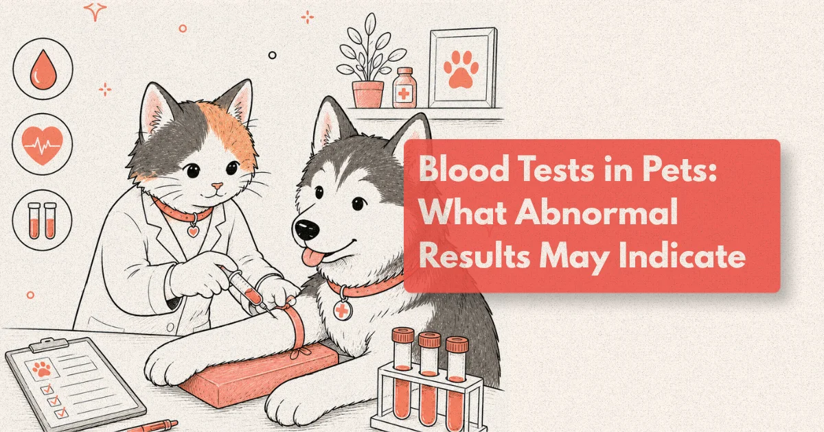
This guide explains the major categories of veterinary blood test, what the key markers measure, and what patterns of abnormality point toward in each organ system.
What to Know
SDMA (symmetric dimethylarginine) detects renal dysfunction when approximately 25% of nephrons are lost, compared to creatinine which requires approximately 75% nephron loss before rising outside the reference range (IRIS, 2023). ALT is elevated in approximately 70% of dogs with active hepatocellular disease at the time of presentation (JVIM, 2022).
How Veterinary Blood Tests Are Structured
A standard veterinary blood panel combines two distinct tests: the complete blood count (CBC) and the serum biochemistry profile. They measure different things and provide complementary information about different aspects of physiology.
The CBC evaluates the cellular components of blood: red blood cells (and their indices including PCV, haemoglobin, MCV, MCHC, and reticulocyte count in anaemic patients), white blood cells and their differential (neutrophils, lymphocytes, monocytes, eosinophils, basophils), and platelets. The CBC answers questions about anaemia, immune response, infection, inflammation, bone marrow function, and clotting capacity.
The serum biochemistry profile measures soluble markers in the liquid fraction of blood after cells are removed. Liver enzymes, kidney markers, proteins, glucose, electrolytes, and pancreatic markers all fall within this category. The biochemistry profile answers questions about organ function, metabolic status, and the integrity of specific tissue compartments.
Additional targeted tests extend the picture when the standard panel raises a specific question. Thyroid hormones for endocrine assessment. Bile acids for functional liver evaluation. Urinalysis combined with urine protein-to-creatinine ratio for refined renal assessment. Cortisol and ACTH stimulation for adrenal disease. Pancreatic lipase immunoreactivity for pancreatitis. Each layer of testing adds specificity to a differential list that the standard panel begins to populate.
The interpretation of any single abnormal value requires context. Reference ranges are established from populations of healthy animals and define the range within which 95% of healthy individuals fall. By definition, 1 in 20 healthy pets will have at least one value outside the reference range on any given panel. A mildly elevated value in an otherwise healthy-looking young animal with no other abnormalities on the panel has a different clinical meaning than the same value in a 12-year-old dog that has been losing weight for two months.
High Liver Values in Dogs: Understanding the Pattern
Liver enzyme elevations are among the most frequently encountered abnormalities on canine biochemistry panels, and they are also among the most commonly misinterpreted. The presence of a high liver value does not indicate liver failure. It indicates that hepatocellular membranes are under stress, that biliary cells are being stimulated, or that an enzyme is being produced in excess by a tissue other than the liver. The clinical significance depends on which enzyme is elevated, by how much, and what accompanies it on the rest of the panel.
The four liver markers reported on standard canine panels are ALT (alanine aminotransferase), ALP (alkaline phosphatase), GGT (gamma-glutamyl transferase), and AST (aspartate aminotransferase). Each has a different tissue origin and different sensitivity for specific types of liver disease.
ALT is highly specific for hepatocellular injury in dogs and cats. It is found in high concentrations within hepatocyte cytoplasm and is released into circulation when hepatocyte membranes are damaged. ALT elevations greater than three times the upper reference limit are generally considered clinically significant and warrant further investigation. Mild ALT elevations (1.5-3x reference) occur with drug administration (non-steroidal anti-inflammatory drugs, phenobarbitone, steroids), strenuous exercise, and non-hepatic disease, and require clinical context for interpretation.
ALP has a wider tissue distribution in dogs than in cats or humans, which makes its interpretation more complex. ALP is produced by hepatocytes, biliary epithelium, bone, intestinal mucosa, and placental tissue. In dogs, a steroid-induced isoform (s-ALP) is produced in response to endogenous or exogenous glucocorticoids, meaning that a dog on prednisolone or one with Cushing’s disease may have ALP values five to twenty times the upper reference limit without primary hepatic disease. Isolated ALP elevation without ALT elevation in a middle-aged to older dog is one of the most common clinical scenarios triggering investigation for hyperadrenocorticism.
A 2023 study of dogs with confirmed hepatocellular carcinoma found that ALT was elevated in 78% at diagnosis, ALP in 85%, and both enzymes together in 71% (Veterinary Comparative Oncology, 2023). The combined pattern is more informative than either marker alone. When both ALT and ALP are elevated at more than three times the reference limit, the probability of primary hepatic disease rises significantly compared to isolated ALP elevation.
Bile acids testing, performed with paired pre-prandial and two-hour post-prandial samples, is the most sensitive functional test for hepatic disease and for portosystemic shunts. Unlike ALT and ALP, which reflect hepatocellular membrane integrity, bile acids measure the liver’s ability to extract and recirculate bile from portal blood. An elevated post-prandial bile acid concentration in a young small-breed dog with any history of neurological signs (seizures, circling, apparent blindness) is a strong indicator for portosystemic shunt and should prompt CT angiography.
From clinical observation: Dogs with steroid-responsive meningitis-arteritis (SRMA) on long-term prednisolone often present with ALP values exceeding 1,000 U/L on routine monitoring panels. Owners understandably find these numbers alarming. In most cases, hepatic ultrasound confirms vacuolar hepatopathy (steroid-induced glycogen accumulation) rather than primary hepatic disease, and the management decision is to optimise the steroid dose rather than investigate the liver independently. This pattern is among the most common sources of unnecessary hepatic referrals.
In-House Laboratory Diagnostics: Speed, Scope, and Limits
In-house laboratory diagnostics refers to blood and urine analysis performed within the veterinary practice using point-of-care (POC) analysers rather than submitted to an external reference laboratory. By 2024, in-house analysers were present in over 90% of companion animal practices across North America, a dramatic shift from the reference-laboratory-only model of a decade earlier (AAHA Practice Survey, 2024). The change has altered the pace of diagnosis in primary care and emergency settings in ways that are clinically meaningful.
The primary advantage of in-house diagnostics is time-to-result. A full biochemistry panel and CBC on an in-house analyser is available in 10-15 minutes. The same panel submitted to an external laboratory returns results in 12-24 hours during standard operating hours, and potentially 48 hours over a weekend or public holiday. In the emergency context, a dog presenting with suspected Addison’s disease, acute pancreatitis, or uroabdomen needs a biochemistry result within the appointment, not the following morning. In-house diagnostics makes same-appointment decision-making possible.
For chronic disease monitoring, the difference between in-house and external laboratory results is more nuanced. Patients with confirmed renal disease, diabetes mellitus, or cardiac disease on digoxin require regular biochemistry monitoring. Running these panels in-house allows the clinician to review and discuss results with the owner during the same appointment, which supports medication adjustment and owner engagement. Serial results are also more directly comparable when run on the same analyser, because inter-analyser variation between platforms can produce apparent shifts that don’t reflect true physiological change.
The limitations of in-house diagnostics are equally important to understand. Haematology accuracy varies by platform and species. In-house CBC analysers perform well for canine samples but have recognised limitations in feline and exotic species haematology, where manual differential cell counts by a trained clinical pathologist add meaningful accuracy. Platelet counting in cats is a known weak point across most POC haematology platforms.
For complex or diagnostically challenging cases, external laboratory submission remains valuable for reasons beyond accuracy. Specialist clinical pathologists at reference laboratories review flagged smears, identify morphological abnormalities (toxic neutrophils, band forms, platelet clumping, abnormal lymphocyte morphology) that POC analysers may not report, and can perform additional tests such as Coombs testing, coagulation panels, and bone marrow cytology on submitted samples. The IDEXX and Antech specialist pathologist review service, available with premium laboratory profiles, adds diagnostic interpretation that in-house printouts cannot replicate.
A 2022 audit comparing in-house and external laboratory results on the same patient samples found that biochemistry values agreed within 10% in 94% of analytes across paired samples, supporting the clinical reliability of modern in-house platforms for routine monitoring (JVIM, 2022). Haematology agreement was lower, particularly for absolute differential white cell counts in cats, which agreed within 15% in approximately 78% of paired samples.
A pattern worth noting: In-house analysers that produce a simultaneous urinalysis (urine specific gravity, dipstick, and sediment) alongside the biochemistry panel add disproportionate value to the renal assessment. A dog with a creatinine of 180 µmol/L (mildly elevated) and a urine specific gravity of 1.048 is concentrating well and likely has pre-renal azotaemia. The same creatinine with a specific gravity of 1.012 (isosthenuric) indicates renal concentrating failure and a far more significant clinical picture. The biochemistry number alone does not tell you which of these patients you are looking at.
Reading a Complete Blood Count
The CBC provides a window into three cell lines: red blood cells, white blood cells, and platelets. Abnormalities in any of these lines carry specific clinical implications that are often more immediately actionable than biochemistry findings.
Anaemia, detected as a low red blood cell count, packed cell volume (PCV), or haemoglobin, is classified as regenerative or non-regenerative based on the reticulocyte count. Regenerative anaemia (high reticulocytes) indicates the bone marrow is responding to blood loss or red blood cell destruction. Non-regenerative anaemia (low or absent reticulocytes) suggests the bone marrow is not responding appropriately, which points toward chronic disease, bone marrow suppression, or primary bone marrow pathology. This distinction changes the differential list and the investigation pathway significantly.
Neutrophilia with a left shift (elevated band neutrophils alongside mature neutrophils) is a classic indicator of bacterial infection or significant tissue inflammation. In a dog presenting with acute abdominal pain and a biochemistry showing elevated BUN and creatinine, neutrophilia with a left shift and toxic neutrophil morphology on the blood smear raises immediate concern for sepsis. In a cat with chronic dental disease and mild weight loss, the same neutrophilia has a much lower urgency rating. Context drives interpretation.
Lymphopenia, a low lymphocyte count, occurs in a range of contexts: stress response (endogenous cortisol release), exogenous steroid administration, viral disease, and lymphoma. Lymphocytosis, conversely, is seen in excitement, chronic antigenic stimulation, and large granular lymphocyte (LGL) lymphoma in cats, a condition associated with alimentary disease and carrying a markedly different prognosis from small cell lymphoma.
Thrombocytopenia (low platelets) below 50 × 10⁹/L carries a bleeding risk. Immune-mediated thrombocytopenia (IMTP) is one of the more common causes in dogs and can present with petechiation, ecchymosis, and mucosal bleeding in the absence of trauma. Tick-borne disease (Ehrlichia, Anaplasma) causes thrombocytopenia as a characteristic finding and should be on the differential list for any dog with unexplained low platelets and appropriate travel or tick exposure history.
Kidney Markers: Creatinine, BUN, and SDMA
Creatinine and blood urea nitrogen (BUN) have been the standard markers for renal function in veterinary panels for decades. Both are waste products that accumulate in the bloodstream when the kidneys fail to clear them at normal efficiency. Both are influenced by non-renal factors that can produce misleading elevations or mask true renal disease.
Creatinine is produced by muscle catabolism at a relatively constant rate. It rises when glomerular filtration rate (GFR) declines significantly, but that significant decline corresponds to approximately 75% of nephron mass being non-functional before creatinine moves outside the reference range (IRIS, 2023). A lean, muscle-wasted cat with chronic kidney disease may have a creatinine value within the reference range despite substantial renal dysfunction, because reduced muscle mass produces less creatinine to accumulate.
SDMA (symmetric dimethylarginine) was introduced to veterinary practice as a more sensitive early marker of renal dysfunction. SDMA is produced by methylation of arginine residues in proteins and excreted almost exclusively by glomerular filtration. It rises when approximately 25% of nephron function is lost, well before creatinine crosses the reference limit. In a 2021 multicentre study, SDMA identified renal dysfunction an average of 17 months earlier than creatinine in cats and 9.5 months earlier in dogs followed longitudinally (JVIM, 2021). That earlier detection creates a larger treatment window for dietary modification, hydration support, and phosphate restriction, all of which slow disease progression.
BUN is less specific than creatinine because it is influenced by dietary protein intake, GI bleeding, and hydration status. A dog fed a high-protein diet will have a higher BUN than the same dog on a renal-support diet, regardless of kidney function. The BUN-to-creatinine ratio is occasionally useful: a ratio above 30:1 in a dog with elevated creatinine suggests pre-renal azotaemia (dehydration reducing kidney perfusion) rather than primary renal failure, and the clinical response to IV fluid therapy confirms or refutes that interpretation.
Thyroid, Pancreatic, and Hormonal Panels
The targeted hormonal and pancreatic tests that extend beyond the standard panel are some of the highest-yield additions in the right clinical context.
Total T4 (thyroxine) is the standard screening test for feline hyperthyroidism, the most common endocrine disorder in cats over ten years of age, affecting approximately 10% of this population (JVIM, 2021). T4 sensitivity for hyperthyroidism in cats with classic clinical signs (weight loss, polyphagia, hyperactivity, palpable thyroid nodule) is approximately 91-98%. The limitation is the approximately 2-10% of hyperthyroid cats with T4 values within the reference range due to concurrent non-thyroidal illness (the “sick euthyroid” phenomenon) suppressing an otherwise elevated T4. Free T4 by equilibrium dialysis has higher sensitivity in these cases and is the appropriate follow-up test when clinical suspicion is high and total T4 is normal.
TSH (thyroid-stimulating hormone) in dogs, measured by cTSH assay, is the primary screening test for hypothyroidism combined with total T4. Low total T4 with elevated cTSH is consistent with primary hypothyroidism. Low T4 with normal cTSH (which occurs in approximately 20% of hypothyroid dogs) requires further evaluation with free T4 and clinical context, because T4 can be suppressed by non-thyroidal illness, obesity, and numerous medications without true thyroid gland disease.
Canine and feline pancreatic lipase immunoreactivity (cPLI, fPLI) are the most specific blood markers for pancreatitis, superseding the older amylase and lipase assays that lacked specificity in both species. Approximately 65-70% of dogs with acute pancreatitis have elevated cPLI at presentation (JVIM, 2022). The sensitivity is higher for moderate-to-severe pancreatitis than for mild disease, and abdominal ultrasound is complementary to PLI for detecting pancreatic changes when PLI is borderline.
Cortisol testing for hyperadrenocorticism (Cushing’s disease) requires dynamic testing rather than a single measurement. The low-dose dexamethasone suppression test (LDDST) is the screening test of choice, with sensitivity of approximately 85-95%. The ACTH stimulation test has lower sensitivity for Cushing’s but is the preferred test for monitoring mitotane (o,p-DDD) or trilostane treatment response. A single resting cortisol below the reference range in a dog with consistent clinical signs (polyuria, polydipsia, bilateral muscle wasting, pot-bellied appearance) is not sufficient to exclude Cushing’s.
Frequently Asked Questions
What does it mean when my dog has high liver enzymes?
Elevated liver enzymes (ALT, ALP, GGT, AST) indicate that hepatocellular membranes are under stress or that biliary cells are being stimulated, but they do not indicate liver failure. ALT elevation is specific to hepatocellular injury; ALP elevation has many causes in dogs, including steroid administration, Cushing’s disease, and primary liver disease. ALT is elevated in approximately 70% of dogs with active hepatocellular disease (JVIM, 2022). The pattern and magnitude of enzyme elevations guide the next investigation step.
Is a single blood test enough to diagnose kidney disease in pets?
A single panel showing elevated creatinine, BUN, and SDMA is strongly suggestive of renal disease but requires urinalysis to interpret. A dog with elevated creatinine and a concentrated urine specific gravity (above 1.030) likely has pre-renal azotaemia from dehydration rather than intrinsic renal disease. The same values with dilute urine (specific gravity below 1.015) indicate renal concentrating failure. SDMA can detect renal dysfunction when only 25% of nephrons are lost, approximately three times earlier than creatinine alone (IRIS, 2023).
Why does my vet recommend both an in-house and external laboratory panel?
In-house panels provide immediate results for same-appointment decisions. External reference laboratory panels add specialist clinical pathologist review of abnormal results, higher accuracy for feline haematology, and access to tests not available on POC analysers (coagulation panels, Coombs testing, Ehrlichia titres, bile acids). For emergency or monitoring work-ups, in-house is appropriate. For complex or diagnostically uncertain cases, external laboratory submission with specialist review adds meaningful interpretive value.
How often should senior pets have blood tests?
Most veterinary guidelines recommend annual bloodwork for healthy pets from age 7, and every 6 months from age 10-11 in dogs or 10 in cats. More frequent monitoring (every 3-6 months) is standard for pets with confirmed organ disease, those on long-term medication (NSAIDs, phenobarbitone, trilostane, chemotherapy), and for monitoring treatment response. SDMA is included in senior panels at most referral centres because of its earlier detection of renal dysfunction compared to creatinine.
Can stress cause abnormal blood test results in pets?
Stress (endogenous cortisol release) causes a predictable pattern on blood tests: mature neutrophilia, lymphopenia, monocytosis, and eosinopenia, termed a “stress leukogram.” Glucose may be mildly elevated in cats with stress hyperglycaemia, which can mimic diabetes mellitus. ALP may be mildly elevated from endogenous glucocorticoid effect. Clinical interpretation requires accounting for the circumstances of blood collection: a fractious cat sampled after a car journey and a waiting room is not in a physiological resting state.
Numbers That Tell a Story
Blood test results are numbers on a page until a clinician places them in context: the age of the patient, the clinical signs that prompted the test, the medications on board, the trend over previous panels, and the organ system where the numbers converge. A mildly elevated ALT in a young Labrador on a monthly flea preventive and a severely elevated ALT in a 12-year-old Cocker Spaniel losing weight describe different situations requiring different responses.
Understanding what each marker measures and which patterns matter most equips owners to engage meaningfully with the clinical conversation rather than respond to the numbers themselves. The number is the beginning of the question; the context is what determines the answer.