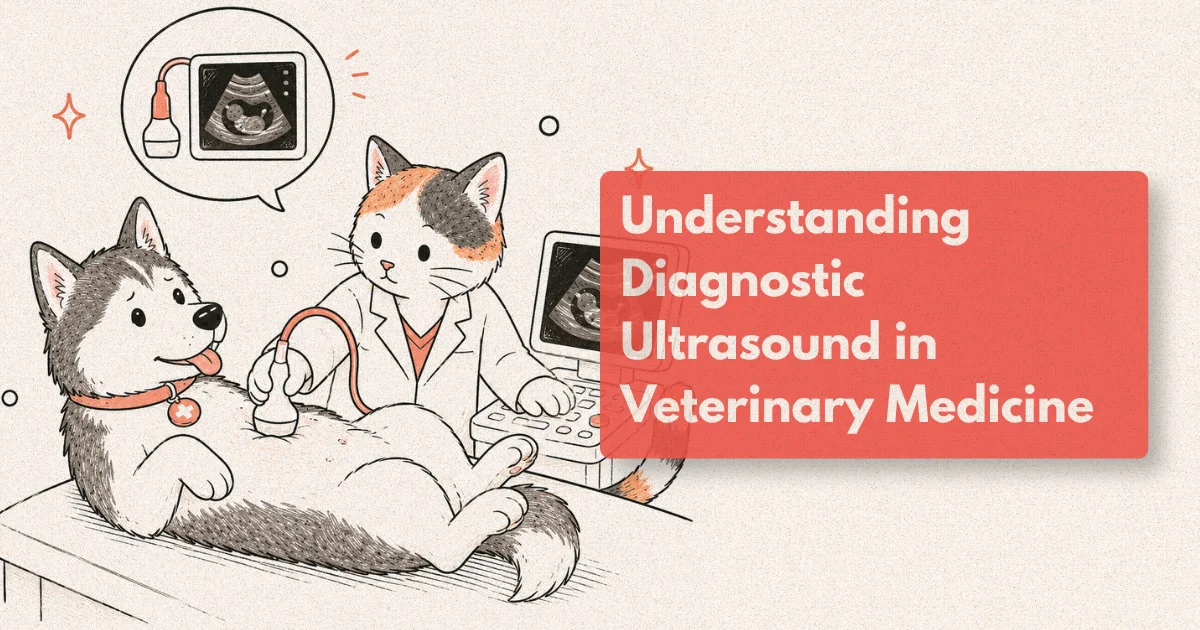
Your veterinarian recommends an ultrasound and your first instinct might be to equate it with pregnancy scans. It’s the same technology, and the physics are identical, but the clinical applications in veterinary medicine are far broader and more varied than that analogy suggests. Ultrasound has become one of the cornerstones of veterinary diagnosis over the past two decades, used in everything from staging a suspected cancer to guiding a needle into a lymph node to monitoring a dog’s heart between annual wellness visits.
What makes it valuable isn’t just what it can find. It’s what it can find without surgery, without radiation, without general anaesthesia in most cases, and in real time. A radiograph shows anatomy frozen in a single moment. Ultrasound shows the liver contracting, a heart valve moving with each beat, a bladder wall with layers you can differentiate, and a mass with internal characteristics that suggest whether it’s benign or malignant. That dynamic quality changes what’s possible in a single outpatient appointment.
This guide explains how the technology works, what each major application actually involves, and what it means for a pet’s diagnostic workup when a veterinarian or specialist recommends it.
What to Know
Diagnostic ultrasound can detect organ abnormalities before bloodwork becomes abnormal in up to 40% of cases, making it a critical first-line imaging tool for pets with non-specific signs like weight loss or lethargy (Journal of Veterinary Internal Medicine, 2023). It’s non-invasive, uses no ionising radiation, and most pets tolerate it without sedation.
How Ultrasound Works in a Clinical Setting
Sound, not radiation, is what makes ultrasound imaging possible. The transducer (the probe the sonographer holds against the patient) emits pulses of high-frequency sound waves, typically in the range of 2-15 MHz depending on the depth and tissue type being evaluated. Those waves travel into the body, reflect off structures with different acoustic properties, and return to the transducer. The system calculates depth from the time it takes each echo to return and builds a real-time image from the reflected data.
Tissues with different densities produce different echogenicity: bone reflects almost all sound (appearing bright white, with a dark shadow behind it), fluid reflects almost none (appearing black), and solid soft tissue falls somewhere between depending on its cell density and composition. This is why a bladder full of urine looks like a clean black circle and a liver with increased fat deposition looks brighter than expected.
The image you see on the screen is updated continuously, typically at 30-60 frames per second, which is what gives ultrasound its real-time quality. A sonographer experienced in veterinary imaging reads the scan as it happens, recognising patterns in echo texture, border regularity, internal architecture, and relative size against reference ranges for the species and breed.
Most abdominal ultrasound examinations in dogs and cats take 30-60 minutes when performed by a specialist. A general practitioner doing a focused scan for a specific question (free fluid in the abdomen, early bladder stone assessment) may complete it in 10-15 minutes. The difference is scope: a specialist performs a systematic survey of all abdominal organs, the sublumbar lymph nodes, the spleen, adrenal glands, and any masses that appear. That thoroughness is why specialist referral changes findings in a meaningful proportion of cases.
Ultrasound for Dogs: Common Clinical Indications
Dogs are referred for abdominal ultrasound for a wider range of reasons than most owners expect. It isn’t reserved for cancer work-ups or serious illness. Chronic vomiting that hasn’t resolved with dietary management, unexplained weight loss in a middle-aged dog, mildly abnormal liver enzymes on a wellness panel, a palpable abdominal mass that needs characterising before a surgical decision: all of these are routine indications.
The liver and spleen are among the structures most consistently evaluated. Hepatic changes ranging from chronic hepatitis to hepatocellular carcinoma to vacuolar hepatopathy all produce identifiable patterns on ultrasound. Splenic nodules are common in older large-breed dogs and carry a wide differential, from benign nodular hyperplasia (the most common cause) to splenic haematoma to mast cell tumour. A 2022 study in Veterinary Radiology and Ultrasound found that splenic nodules under 2 cm in dogs had a benign histological diagnosis in approximately 70% of cases, a figure that provides useful context when owners face the decision of whether to monitor or intervene (Veterinary Radiology and Ultrasound, 2022).
Urinary tract assessment is another high-frequency application. Bladder wall thickness, sediment, polyps, and masses are all well visualised. Upper urinary tract imaging, covering the kidneys, renal pelvis, and proximal ureters, is particularly useful for dogs with suspected pyelonephritis or ureteral obstruction from stones or a mass. Hydronephrosis, the dilation of the renal collecting system from obstruction, shows up clearly as a fluid-filled expansion of structures that should be compact.
For dogs with suspected adrenal disease, Cushing’s syndrome being the most common presentation, ultrasound helps distinguish pituitary-dependent disease (typically bilateral adrenal enlargement) from adrenal-dependent disease (a unilateral adrenal mass, often with the contralateral gland appearing atrophied). This distinction directly influences treatment planning and whether surgical adrenalectomy is on the table.
From clinical observation: Dogs with mild, chronic GI signs are among the most frequently referred for abdominal ultrasound in internal medicine settings. What often appears to be a routine dietary intolerance case turns out, on ultrasound, to have diffuse intestinal wall thickening consistent with inflammatory bowel disease or alimentary lymphoma, diagnoses that require biopsy confirmation but that the ultrasound identifies as the priority. Bloodwork in these dogs is often only mildly abnormal or entirely normal.
What Ultrasound Imaging Actually Reveals
Reading a veterinary ultrasound image isn’t a matter of spotting obvious tumours. Most of what a trained sonographer assesses is subtle: the echogenicity of a parenchymal organ relative to its neighbours, the layered wall architecture of the intestine, the smoothness of a kidney’s cortical margin, the presence or absence of portal vein flow on Doppler interrogation. The technique requires pattern recognition built from thousands of examinations, which is why a board-certified veterinary radiologist or internal medicine specialist produces a qualitatively different report than a general practitioner using the same machine.
The intestinal wall is one of the more information-rich structures on abdominal ultrasound. A healthy small intestinal wall in a dog has five visible layers: the outer serosa (hyperechoic), the muscularis (hypoechoic), the submucosa (hyperechoic), the mucosa (hypoechoic), and the mucosal surface (hyperechoic). When those layers lose definition or one layer becomes disproportionately thickened, it’s meaningful. Diffuse mucosal thickening without loss of layering suggests inflammatory disease. Loss of the normal layering pattern raises concern for neoplasia, particularly lymphoma. This distinction guides the biopsy approach: endoscopy or full-thickness surgical biopsy depending on the depth and distribution of change.
Lymph node assessment is another dimension with significant clinical weight. Normal sublumbar and mesenteric lymph nodes are small and fusiform. Rounded, enlarged lymph nodes with a loss of the normal fatty hilus are a consistent ultrasound finding in lymphoma and metastatic disease. A 2021 study in the Journal of Veterinary Internal Medicine found that lymph node short-axis measurement greater than 1 cm in mesenteric nodes had a sensitivity of 82% and specificity of 79% for distinguishing reactive from neoplastic disease in dogs, a figure that supports using it as a triage criterion for cytology sampling (JVIM, 2021).
Ultrasound-guided sampling is where imaging and diagnosis converge most directly. Fine needle aspiration (FNA) of an ultrasound-identified mass or lymph node, performed with the needle tip visualised in real time as it enters the target, transforms what would otherwise require exploratory surgery into a same-day outpatient procedure. In dogs, ultrasound-guided FNA yields diagnostic cytology in approximately 75-80% of focal hepatic lesions when performed by an experienced operator (Veterinary Radiology and Ultrasound, 2020). For cases where cytology is insufficient and a tissue core is needed, ultrasound-guided Tru-Cut biopsy provides histological material without an incision.
Doppler ultrasound adds a vascular layer to the structural picture. Colour flow Doppler shows whether a mass has internal vascularity (more consistent with malignancy) versus appearing avascular (more consistent with a simple cyst or haematoma). Portal vein flow assessment helps characterise hepatic disease and identify portosystemic shunts, particularly in young small-breed dogs presenting with neurological signs, failure to thrive, or post-prandial hypoglycaemia.
A pattern worth noting clinically: Practitioners sometimes encounter a dog where the abdominal ultrasound appears unremarkable, yet the clinical signs are clearly GI. In these cases, the normal ultrasound is itself informative: it reduces the probability of structural disease and shifts the differential toward functional or mucosal disorders that are below ultrasound resolution, guiding the next step toward endoscopy rather than surgery.
Ultrasound in Veterinary Internal Medicine
Veterinary internal medicine is the specialty that most frequently orders, interprets, and acts on ultrasound findings in a comprehensive way. A board-certified specialist in veterinary internal medicine (Diplomate, ACVIM) uses ultrasound as a clinical tool within a broader diagnostic framework that includes history, physical examination, laboratory data, and endoscopy when indicated. The ultrasound is rarely the only test. It’s the test that contextualises everything else.
In 2024, there were approximately 2,800 ACVIM Diplomates practicing across all internal medicine specialties in North America, a 35% increase from a decade earlier (ACVIM, 2024). Most maintain ultrasound proficiency as a core clinical skill, and many have completed additional training in advanced sonographic techniques including contrast-enhanced ultrasound (CEUS) and elastography.
Endocrine disease is one of the areas where the internal medicine and imaging disciplines intersect most productively. Hypoadrenocorticism (Addison’s disease), hypothyroidism, hyperadrenocorticism (Cushing’s), and diabetes mellitus all have ultrasound correlates. Adrenal glands that are bilaterally small (under 3.5 mm craniocaudal diameter) in a dog with suspected Addison’s support that diagnosis. Asymmetric adrenal enlargement in a Cushing’s patient shifts the probability toward adrenal-dependent disease and prompts cross-sectional imaging for surgical planning.
Protein-losing enteropathy (PLE), one of the more challenging conditions in small animal internal medicine, benefits significantly from systematic ultrasound assessment. Diffuse intestinal wall changes, lymphangiectasia visible as hyperechoic foci in the intestinal mucosa, hypoechoic mesenteric fat, and free abdominal fluid (ascites from hypoalbuminaemia) are collectively an ultrasound picture that supports PLE before invasive testing confirms it. A 2022 retrospective at a tertiary referral centre found that ultrasound identified a contributory abnormality in 83% of dogs ultimately diagnosed with PLE (JVIM, 2022).
Point-of-care ultrasound (POCUS) has extended ultrasound use into emergency and critical care settings where specialist sonographers aren’t always present. Emergency clinicians trained in the AFAST (Abdominal Focused Assessment with Sonography for Trauma) and TFAST (Thoracic FAST) protocols can identify free fluid, pericardial effusion, and pneumothorax within minutes at the bedside. This capability has changed triage decision-making in ways that are clinically meaningful: a dog in haemorrhagic shock with a positive AFAST (free peritoneal fluid visible in standard scan windows) moves directly to surgery rather than waiting for a radiograph and interpretation.
A prospective study found that POCUS-positive AFAST findings in trauma patients predicted the need for emergency surgery with a sensitivity of 88% and specificity of 86%, enabling surgical intervention significantly faster than the conventional diagnostic pathway (Lisciandro, Veterinary Emergency and Critical Care, 2011; replicated in multicentre data 2022). This shift from a radiograph-dependent to a sonography-first approach is now standard at most 24/7 emergency facilities.
What Happens During Your Pet’s Ultrasound Appointment
Knowing what to expect reduces the anxiety around the appointment itself. Most abdominal ultrasounds are performed in a padded V-trough or on a table with the patient in dorsal or lateral recumbency. The abdomen is clipped (fur blocks acoustic contact) and ultrasound gel is applied to improve transducer-skin coupling. Most dogs and cats tolerate this without sedation. The scanning itself is painless.
For a comprehensive abdominal study, the sonographer works through the organ systems systematically: liver, gallbladder and bile duct, stomach, small intestine, large intestine, spleen, kidneys, adrenal glands, urinary bladder, and regional lymph nodes. Measurements are taken of organs that are abnormal in size. Any masses, fluid pockets, or unexpected findings are documented with still images and cine clips.
Sedation is occasionally used for anxious patients or when a precise guided sample is planned, but it isn’t routine for straightforward abdominal assessment. Thoracic ultrasound, including echocardiography, is usually performed with the patient standing or in lateral recumbency, with the probe applied through the intercostal spaces. Cardiac studies take longer and require more patient cooperation than abdominal studies.
When an ultrasound-guided FNA or biopsy is performed at the same appointment, the area is clipped and cleaned. Local anaesthetic may be injected into the skin. The needle is introduced through the body wall under real-time guidance while the patient is awake or lightly sedated. The procedure takes minutes. Most patients show no more than brief discomfort.
Results from the imaging interpretation itself are typically available the same day from the performing clinician. If samples were collected for cytology or histopathology, results take 24-72 hours from an external laboratory. The internal medicine or radiology team usually provides a written report that your general practitioner integrates into the broader management plan.
Echocardiography: The Heart Under the Probe
Echocardiography is ultrasound focused entirely on the heart: its structure, dimensions, wall motion, valve function, and blood flow dynamics. It’s the definitive diagnostic test for cardiac disease in dogs and cats and is used both for diagnosis at clinical presentation and for screening in breeds with known heritable cardiac conditions.
Cavalier King Charles Spaniels are the most studied example. The ACVIM consensus guidelines recommend annual echocardiographic screening beginning at age 2, with stricter protocols for breeding animals, because mitral valve disease is heritable and detectable before clinical signs appear (ACVIM Consensus Statement on MVD, 2019). Maine Coons and Ragdolls undergo echocardiographic screening for hypertrophic cardiomyopathy (HCM) for the same reason: the genetic mutations are known, the disease is detectable in the preclinical phase, and early intervention with medication changes the timeline.
A standard echocardiographic study in a dog or cat measures left ventricular internal dimensions in systole and diastole, wall thickness, fractional shortening (a measure of contractile function), and valve morphology. Doppler interrogation quantifies regurgitant flow velocity, which allows grading of valve disease severity. In cats, diastolic function is assessed through transmitral flow patterns and tissue Doppler imaging, because HCM affects the heart’s ability to relax and fill rather than its ability to contract.
Cardiac output, ejection fraction, and estimated pulmonary arterial pressure (via tricuspid regurgitation jet velocity on continuous-wave Doppler) round out the functional picture. These numbers guide medication decisions, follow-up interval, and prognosis conversations with the owner.
Frequently Asked Questions
Do dogs need to be sedated for an abdominal ultrasound?
Most dogs don’t require sedation for a routine abdominal ultrasound. The procedure is painless and non-invasive, though the abdomen does need to be clipped and gel applied. Sedation is reserved for anxious patients or when ultrasound-guided sampling is planned alongside the scan. Cats are more variable; some tolerate it readily while others benefit from mild anxiolytic sedation (ACVR, 2023).
What can a veterinary ultrasound detect that bloodwork cannot?
Structural changes in organ size, shape, internal architecture, and the presence of masses or fluid are invisible to bloodwork. Ultrasound detects organ abnormalities before bloodwork turns abnormal in up to 40% of cases (JVIM, 2023). Bloodwork reflects systemic function; ultrasound reveals structural anatomy. The two complement each other, and neither alone gives the full picture.
How accurate is ultrasound for detecting cancer in pets?
It depends on the organ and lesion type. Ultrasound-guided FNA achieves diagnostic cytology in 75-85% of hepatic and lymph node lesions in experienced hands (Veterinary Radiology and Ultrasound, 2020, 2022). Splenic and adrenal masses have lower cytological yield (55-65%), and in those cases a Tru-Cut biopsy or surgical removal may be needed for definitive diagnosis. Ultrasound finds the lesion; biopsy confirms what it is.
What’s the difference between a general vet doing an ultrasound and a specialist?
A board-certified veterinary radiologist or internal medicine specialist performs a systematic, comprehensive survey of all abdominal structures with specialist-level pattern recognition. Studies comparing general practitioner and specialist ultrasound reports on the same patient have found that specialists identify additional clinically significant findings in 30-40% of cases (Veterinary Radiology and Ultrasound, 2021). For complex or high-stakes diagnostic questions, specialist ultrasound is worth the referral.
How often should dogs with known heart disease have echocardiograms?
It depends on the disease stage. Dogs in the preclinical phase of mitral valve disease (Stage B1 under ACVIM guidelines) are typically reassessed every 12 months. Those in Stage B2 (enlarged heart, still asymptomatic) are started on medication and reassessed every 6 months. Dogs in heart failure (Stage C) may have echocardiograms every 3-6 months to monitor medication response (ACVIM MVD Consensus Statement, 2019).
The Image That Changes the Plan
Diagnostic ultrasound doesn’t replace clinical judgment. It extends it. The most useful description of what ultrasound does in a veterinary workup is that it converts a clinical suspicion into a structured, anatomically located question: is this liver change diffuse or focal? Is this mass vascular? Is there free fluid in the abdomen? Once those questions have answers, the path from diagnosis to treatment becomes substantially cleaner.
The technology has become democratised enough that general practitioners use it daily for focused questions, while specialists use it as the foundation of complex internal medicine work-ups. Whether your dog is having a lump evaluated or your cat is being screened for a heritable heart condition, the probe and the physics underneath it are doing the same thing: turning sound into information.