When a dog has been vomiting for three weeks with normal bloodwork and a non-diagnostic abdominal ultrasound, or when a cat develops progressive hindlimb weakness that doesn’t localise cleanly on neurological examination, the conversation eventually turns to CT or MRI. Advanced imaging doesn’t replace the diagnostic steps that came before it. It extends them into anatomical territory that radiography and ultrasound can’t reach with the precision the clinical question requires.
CT (computed tomography) and MRI (magnetic resonance imaging) both require general anaesthesia in veterinary patients because the acquisition demands complete stillness that cannot be achieved in an awake animal. That requirement introduces cost and logistical complexity. It also means the decision to pursue advanced imaging involves a genuine risk-benefit conversation, not just a financial one. When that conversation is well-informed, patients reach the right answer faster and with fewer redundant procedures along the way.
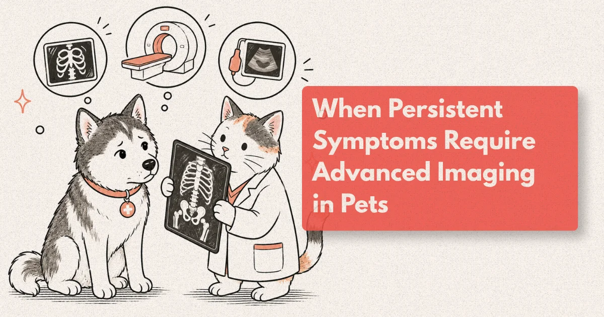
This guide explains the clinical reasoning behind advanced imaging referrals, what each modality actually shows, and what the experience of an imaging appointment involves for pets and owners navigating it for the first time.
What to Know
CT detects pulmonary nodules as small as 1-2 mm, compared to the 8-10 mm detection threshold of standard radiography (Journal of Veterinary Internal Medicine, 2022). In dogs with nasal disease, CT changed the clinical management decision in 73% of cases compared to radiography alone (Veterinary Radiology and Ultrasound, 2019). General anaesthesia is required for both CT and MRI in veterinary patients.
When Standard Diagnostics Are Not Enough
The referral for advanced imaging typically follows a recognisable pattern: clinical signs are present, they’re significant enough to pursue, and the available diagnostics have either pointed toward a region that can’t be assessed at sufficient resolution or returned results that don’t match the clinical picture. Radiography and ultrasound are excellent first-line tools. They’re also tools with specific resolution ceilings.
Radiography produces a two-dimensional summation of three-dimensional anatomy, with overlapping structures that obscure fine detail and limit the ability to characterise soft-tissue lesions. Ultrasound delivers excellent real-time soft-tissue information but loses fidelity where bone blocks sound transmission (the skull, the vertebral canal, the thoracic cavity) and where gas-filled structures scatter the beam (the lung parenchyma, portions of the gastrointestinal tract). These limitations are inherent to the physics, not shortcomings in the technique.
Neurological signs that localise to the brain or spinal cord are among the most consistent indications for advanced imaging. A dog with acute-onset seizures, progressive ataxia, or unexplained cranial nerve deficits requires intracranial imaging. A cat with progressive paraparesis needs spinal MRI or CT myelography. These patients cannot be managed appropriately without knowing whether the lesion is compressive (surgery may be indicated) or parenchymal and inflammatory (medical management is the primary approach). A neurological examination may localise the lesion to a spinal cord segment, but it cannot tell you what the lesion is.
Nasal and paranasal sinus disease is another category where radiography systematically under-performs. CT of the nasal cavity and skull base shows tumour extent, cribiform plate involvement, orbital invasion, and regional lymph node status in a single acquisition. Thoracic staging for confirmed malignancy, vascular anomalies such as portosystemic shunts, complex orthopaedic injuries, and pre-surgical planning for adrenal or hepatic masses complete the most common indications across species and age groups.
CT Scan for Dogs: What the Technology Shows and When It Matters
CT works by rotating an X-ray source and detector array around the patient, acquiring cross-sectional data that a computer reconstructs into three-dimensional volumetric images. Modern multidetector CT (MDCT) scanners complete thoracic and abdominal acquisition in under 60 seconds of table time, which meaningfully reduces the total duration of anaesthesia compared to older single-detector units (Veterinary Radiology and Ultrasound, 2020). The resulting image sets can be reviewed in any anatomical plane, at any slice thickness, and rendered as three-dimensional surface models for surgical planning.
For dogs specifically, the most clinically impactful applications are thoracic staging, nasal and skull imaging, portosystemic shunt characterisation, and pre-surgical planning for complex masses.
Thoracic staging for malignant disease is among the highest-volume CT applications. CT detects pulmonary nodules as small as 1-2 mm, compared to the 8-10 mm practical detection floor for thoracic radiography (JVIM, 2022). When a soft-tissue sarcoma has been removed from a dog’s limb and the clinical question is whether there is pulmonary metastatic disease affecting prognosis, a radiograph that reports clear lungs and a CT that identifies five sub-centimetre pulmonary nodules represent fundamentally different clinical situations. That distinction changes whether adjuvant chemotherapy is recommended and what the owner understands about the prognosis.
Nasal disease illustrates the decision-changing power of CT most clearly. A dog with unilateral epistaxis, chronic nasal discharge, and facial swelling may have nasal lymphoma, nasal carcinoma, aspergillosis, or a foreign body. CT differentiates these by showing the pattern of bone destruction, the extent of soft-tissue infiltration, and whether the cribiform plate separating the nasal cavity from the brain has been crossed. Approximately 70% of nasal tumours that appear to involve the cribiform plate on CT are confirmed inoperable without exploratory surgery (Veterinary Radiology and Ultrasound, 2021). The CT tells the surgeon whether the procedure is worthwhile before anyone is anaesthetised for it.
For portosystemic shunts, CT angiography detects and anatomically characterises shunts with sensitivity exceeding 95%, identifying the exact vessel anatomy before surgical ligation and enabling the surgeon to plan the approach and anticipate all contributing vessels (JVIM, 2021). This is a meaningful advance over ultrasound Doppler, which detects shunts with sensitivity of approximately 70-85% in experienced hands but cannot always delineate shunt type and location with the precision needed for surgical planning.
From clinical observation: Dogs referred for CT with a suspected portosystemic shunt often arrive after a prolonged diagnostic journey involving multiple blood tests, dietary modifications, and inconclusive ultrasound reports. CT angiography in a single anaesthetic episode routinely provides the shunt anatomy, confirms surgical candidacy, and enables scheduling of the corrective procedure. The diagnostic compaction is one of the most clinically satisfying aspects of the modality.
What CT Imaging Reveals at the Tissue Level
The core clinical value of CT imaging is the simultaneous visualisation of bone, soft tissue, and vascular structures in the same acquisition, at sub-millimetre resolution, without the superimposition artefact that degrades radiographic interpretation. That combination makes CT the modality of choice for any clinical question involving the interface between bone and adjacent soft tissue, or between a mass and its surrounding vascular supply.
Contrast-enhanced CT (CE-CT) adds a vascular dimension that transforms mass characterisation. Iodinated contrast is administered intravenously and images are acquired at defined intervals after injection: the arterial phase captures vessels at peak contrast concentration; the portal venous phase captures the liver and spleen parenchyma; the delayed phase reveals slow-enhancing structures such as fibrotic lesions. This three-phase acquisition is standard for hepatic mass staging and for identifying the vascular supply of large tumours before surgery, where knowing whether a mass encases the caudal vena cava changes the surgical risk calculation entirely.
Dynamic airway imaging is another domain where CT has displaced other modalities. In dogs with suspected tracheal collapse, inspiratory and expiratory CT reveals the dynamic reduction in tracheal lumen that no static radiograph can capture during the natural breathing cycle. A prospective study found inspiratory-expiratory CT had a sensitivity of approximately 90% for detecting clinically significant tracheal collapse, compared to approximately 65% for fluoroscopy in the same patient cohort (Veterinary Radiology and Ultrasound, 2022).
CT-guided tissue sampling extends the diagnostic reach into regions that were previously accessible only through surgery. A needle advanced under real-time CT visualisation can reach a target in the thoracic mediastinum, the retroperitoneum, or the lung parenchyma with sub-centimetre precision. CT-guided biopsy avoids exploratory surgery in approximately 65-75% of thoracic mass cases by yielding diagnostic histopathology in a single outpatient procedure (Veterinary Radiology and Ultrasound, 2020).
A multicentre retrospective comparing CT-guided and ultrasound-guided biopsy of thoracic lesions found CT guidance achieved diagnostic histopathology in 71% of cases where a prior ultrasound-guided attempt had been non-diagnostic. The authors attributed this to superior needle-to-target visualisation and avoidance of overlying aerated lung, which scatters the ultrasound beam and degrades real-time guidance. (Veterinary Radiology and Ultrasound, 2020.)
A pattern worth recognising: CT studies read without the clinical context sometimes generate long differential lists that aren’t clinically useful. The most actionable CT reports are written by specialists who have spoken with the referring clinician before the appointment: they know whether the dog’s lymph nodes are the primary question or the incidental finding, and they frame their interpretation accordingly.
Specialist Diagnostics: Who Reads the Images and Why It Changes the Report
Advanced imaging in veterinary medicine is interpreted by board-certified specialists in veterinary radiology (Diplomate, ACVR) or by boarded clinicians in the relevant discipline who have acquired advanced imaging competency during residency training. The distinction between a machine that produces images and a specialist who interprets them is not trivial.
There were approximately 500 ACVR Diplomates in active clinical practice in North America as of 2024, across the diagnostic imaging, radiation oncology, and nuclear medicine tracks (ACVR, 2024). The diagnostic imaging track produces the radiologists who read CT and MRI studies in referral hospital settings. Their training involves a minimum three-year residency following an internship, with case-load requirements covering thousands of multimodality studies across species. Pattern recognition at this level cannot be replicated by a clinician who uses CT occasionally.
When a general practitioner refers a patient for CT, the specialist performs more than image acquisition. They design the protocol before the appointment (which contrast phases, which window widths, whether three-dimensional reconstruction is needed for surgical planning), supervise the anaesthetic event, interpret the study against the clinical history provided by the referring clinician, and produce a written report listing findings, differential diagnoses ranked by probability, and recommended next steps. The diagnostic quality of the report reflects the specialist’s clinical reasoning as much as the machine’s resolution.
Studies comparing general practitioner and specialist interpretation of the same imaging studies found that specialists identified additional clinically significant findings or changed the principal diagnosis in 30-40% of cases (Veterinary Radiology and Ultrasound, 2021). This is not a criticism of general practitioners but a reflection of the specialised training and case volume that radiology training instils. The same effect exists in human medicine, where subspecialist radiology reads have measurably higher sensitivity and specificity for focal diseases than general radiologists in several published comparisons.
For owners, the practical implication is that a referral for advanced imaging is not simply a request to use a bigger machine. It is an engagement with a diagnostic specialist whose professional training is built entirely around turning complex multimodality imaging data into actionable clinical conclusions. Choosing a referral centre where the radiologist is accessible to the clinician for discussion before and after the study adds value beyond the written report alone.
MRI versus CT: Matching the Modality to the Clinical Question
The comparison between MRI and CT is best understood as a difference in contrast mechanism rather than a capability hierarchy. CT uses differential X-ray attenuation to distinguish tissues. MRI uses the behaviour of hydrogen nuclei in a magnetic field to generate contrast between tissues with different water content, fat content, and molecular environment. MRI sees soft-tissue differences that CT cannot resolve. CT sees bone and calcification detail that MRI blurs. Neither is superior in the abstract; each is superior for specific clinical questions.
For the brain and spinal cord, MRI is the standard of care. MRI detects intramedullary spinal cord lesions (within the cord substance itself) with sensitivity approaching 95%, compared to approximately 60% for CT myelography in controlled comparisons (JVIM, 2022). It characterises lesion signal intensity in ways that help distinguish inflammatory conditions from neoplasia from vascular events, guiding the question of whether biopsy, medical management, or radiation therapy is the appropriate next step. For intracranial disease in dogs and cats, MRI is the modality that most directly influences treatment planning.
For thorax, abdomen, nasal cavity, and orthopaedics, CT is the workhorse. Its speed advantage is decisive in patients where prolonged anaesthesia carries meaningful risk: a thoracic and abdominal CT takes under two minutes of acquisition time, while an MRI study of the same region takes 30-90 minutes. Superior bone detail is essential for nasal, skull base, and vertebral cases. Wide availability at specialist centres means the clinical wait time for CT is typically shorter than for MRI at most referral hospitals.
The choice between the two modalities is a question of which one answers the specific clinical question with the highest diagnostic precision at the lowest necessary risk. A well-framed referral question, communicated by the general practitioner to the specialist before the appointment, is what ensures the right modality is selected and the protocol is designed to answer it.
What Happens at the Imaging Appointment
Pets arriving for CT or MRI are assessed by an anaesthesiologist or anaesthesia-trained specialist before induction. Pre-anaesthetic bloodwork, if not already current, is typically recommended for patients over seven years of age or those with known systemic disease. In healthy dogs, the risk of anaesthetic mortality across all elective procedures is approximately 1 in 1,800 (Brodbelt et al., JAVMA, 2008). Risk increases with age, pre-existing organ disease, and emergency presentation, which is why the pre-anaesthetic assessment is not a formality.
Fasting is required: typically 8-12 hours for food and 2-4 hours for water, depending on the facility and anaesthetic protocol. On arrival, the patient is weighed, an intravenous catheter is placed, and induction is performed with short-acting agents. For CT, the anaesthetic episode is generally 30-60 minutes including induction, positioning, scan acquisition, and early recovery. For MRI, the total anaesthetic time is longer, typically 60-120 minutes depending on region and number of sequences.
Contrast agent administration, where indicated, adds a few minutes to the procedure. Iodinated contrast for CT and gadolinium-based contrast for MRI are both generally well tolerated in patients without pre-existing renal disease, though pre-treatment hydration and renal function assessment are standard precautions at most centres.
Results from the imaging interpretation are typically available on the same day or by the following morning as a written report transmitted to the referring veterinarian. If CT-guided sampling was performed during the same anaesthetic episode, cytology or histopathology results follow from the external laboratory within 24-72 hours. The specialist team usually communicates the key imaging findings to the referring clinician verbally before the written report is finalised, enabling same-day management decisions in time-sensitive cases.
Frequently Asked Questions
Does my pet need to be fully anaesthetised for CT or MRI?
Yes. General anaesthesia is required for both CT and MRI in dogs and cats. The acquisition requires complete stillness for the duration of scanning, which cannot be achieved through sedation alone. In healthy dogs, anaesthetic mortality risk across all elective procedures is approximately 1 in 1,800 (Brodbelt et al., JAVMA, 2008). Risk increases with age and pre-existing disease, which is why a pre-anaesthetic health assessment is performed before every imaging appointment.
How long does a veterinary CT scan take?
The scan acquisition itself takes under 2 minutes for most thoracic and abdominal studies on modern MDCT equipment. Including induction, positioning, contrast administration, and early recovery, the total anaesthetic episode is typically 30-60 minutes. Studies requiring multiple contrast phases or three-dimensional reconstruction add modest time. MRI studies take significantly longer: 45-120 minutes depending on anatomical region and number of sequences.
What should I tell the specialist before the CT appointment?
A concise clinical summary improves the study: presenting signs and duration, all medications including supplements, previous diagnostics and results, and the specific question you need the imaging to answer. If prior imaging exists, send it digitally before the appointment. Specialists use this information to design the protocol before the patient arrives, which avoids acquiring images that don’t answer the clinical question and reduces anaesthetic time.
Will the CT result give us a definitive diagnosis?
CT characterises lesions but rarely provides a definitive tissue diagnosis on its own. A hepatic mass on CT generates a differential list. CT narrows it by showing size, location, vascular involvement, and enhancement pattern. Definitive diagnosis usually requires cytology or histopathology, either from CT-guided sampling during the same anaesthetic episode or from surgery. The CT result guides that decision and gives the surgeon or oncologist the structural information they need before a needle or scalpel is used.
Is CT available at my regular vet or do I need a specialist referral?
CT is available primarily at specialist referral centres and veterinary teaching hospitals, though a small number of larger general practices have installed units in recent years. Interpretation expertise is distinct from machine access: when CT is available at a general practice, images are often read by a remote ACVR radiologist via teleradiology services. For complex or high-stakes diagnostic questions, an in-person specialist consultation before and after imaging adds diagnostic value that a remote read alone cannot fully replicate.
The Question That Earns the Anaesthetic
Advanced imaging is not a first step. It’s a precise answer to a precise question, reached after simpler tools have given what they can. The decision to proceed rests on whether the clinical question requires the resolution that CT or MRI provides, whether the patient’s anaesthetic risk is acceptable, and whether the result will change what happens next. When all three conditions are met, the scan that follows tends to move the case forward in ways that weeks of inconclusive blood tests cannot.
The technology itself is secondary. A CT machine in the hands of a general practice without specialist radiological interpretation is a less capable tool than the same machine at a referral centre where a Diplomate reads every study and speaks with the referring clinician about the results. The diagnostic quality of advanced imaging is, in the end, a function of the specialist behind the report.