The rabbit occupies an unusual position in veterinary medicine: it is one of the most commonly kept small mammals as a companion animal and simultaneously one of the species most likely to be seen by a practitioner with limited exotic pet experience. The consequences of that gap are significant. The two most important disease categories in pet rabbits, gastrointestinal stasis and dental disease, are both conditions where the window between early signs and life-threatening deterioration is measured in hours to days rather than weeks, and where the clinical approach differs substantially from that of the dog and cat.
Understanding these conditions properly begins with the rabbit’s physiology, because both diseases are ultimately physiological consequences of the same set of conditions: a digestive system designed for continuous, high-volume fibre processing that is disrupted by diet, environment, or structural disease. The gut and the dentition in the rabbit are functionally inseparable, and the clinician who treats one without evaluating the other is likely to be managing consequences rather than causes.
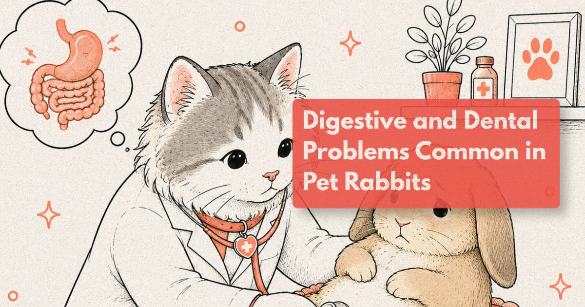
What to Know
Gastrointestinal stasis is estimated to be the most common reason pet rabbits are presented as emergencies in small exotic practice. Studies of rabbit dental disease indicate that over 70% of rabbits presented for any health concern show radiographic evidence of dental abnormality when skull imaging is performed (Harcourt-Brown, 2006). Rabbit stopped eating is never a safe wait-and-see presentation: it is a sign of serious, potentially life-threatening disease until proven otherwise.
The Gut That Never Stops: Why Rabbit Digestive Anatomy Is Unforgiving
The rabbit is an obligate hindgut fermenter. Its digestive strategy is built around continuous intake of high-fibre plant material, rapid passage of indigestible fibre through the stomach and small intestine to the large intestine and caecum, and prolonged fermentation of the resulting substrate in the caecum by a specialised microbial population. This produces not only energy and volatile fatty acids but also caecotropes, nutrient-rich, soft faecal pellets coated in mucus that the rabbit consumes directly from the anus, primarily at night. Caecotrophy is not a behavioural oddity: it is an essential part of the rabbit’s nutrient cycle, providing B vitamins, microbial protein, and additional fermentation products that the primary digestive pass does not capture.
The clinical consequence of this physiology is that gut motility is not optional in the rabbit. Normal peristaltic movement through the stomach and intestines depends on the continuous mechanical stimulation that dietary fibre provides. When fibre intake drops, because the diet is inadequate, because the rabbit is in pain and not eating, or because dental disease makes eating painful, gut motility slows. A slowing gut does not empty properly. Ingesta and gas accumulate. The caecal microbiome, deprived of its normal substrate, shifts toward potentially harmful bacterial populations. The rabbit becomes increasingly uncomfortable, increasingly reluctant to eat, and increasingly compromised in a cycle that accelerates its own deterioration if not interrupted by clinical intervention.
This is the mechanism of gastrointestinal stasis, and understanding it makes clear why the standard approach of waiting 12 to 24 hours to see if a rabbit that has stopped eating “improves” is clinically indefensible. A rabbit that has not eaten for six to eight hours requires veterinary assessment. A rabbit that has not eaten for 12 hours is already at risk of hepatic lipidosis.
Rabbit Stopped Eating: Recognising GI Stasis Before It Becomes Critical
Gastrointestinal stasis is a clinical sign, not a diagnosis. When a rabbit stops eating, the practitioner’s task is not to confirm stasis but to identify what is causing it. Stasis is the final common pathway for a wide range of underlying conditions, and treating it symptomatically without addressing the cause produces temporary improvement at best and allows progression of serious underlying disease at worst.
The most important causes of GI stasis in pet rabbits are dental disease, low dietary fibre, pain from any source, acute stress, environmental change, and dehydration. In a significant proportion of cases, more than one factor is present simultaneously. Dental disease deserves emphasis here because it is both the most common identifiable underlying cause and the most commonly missed: a rabbit that presents in stasis will very often have had dental disease progressing silently for weeks or months before the anorexia became apparent to the owner.
Clinical signs of GI stasis in approximate progression are: reduced food intake (owners often notice the hay rack is less depleted), reduced faecal output or smaller than normal faecal pellets, hunched posture, bruxism (tooth grinding, a pain indicator in rabbits), and, in progressive cases, abdominal distension as gas accumulates. A silent, distended abdomen is an emergency: gas accumulation in the caecum or intestines produces rapidly escalating pain and can progress to ischaemia.
Assessment in the stasis rabbit includes abdominal palpation for gas, distension, or any discrete mass; auscultation of gut sounds (normal rabbit gut is audibly active; silence is a clinical finding); body weight relative to previous records; body temperature (stasis rabbits are often hypothermic due to pain and reduced metabolic activity); and, critically, a thorough oral examination. The limitations of awake oral examination in rabbits are real, and the cheek teeth cannot be adequately assessed without sedation, but the incisors and the rostral cheek teeth can be evaluated at conscious examination, and any obvious malocclusion, incisor elongation, or wet dewlap from hypersalivation is immediately informative.
From clinical practice: One of the most instructive GI stasis presentations is the rabbit referred after two to three weeks of “intermittent” reduced appetite, treated empirically with gut motility agents and syringe feeding, showing partial and temporary improvement between episodes. When the actual cause is dental disease, this pattern is almost diagnostic: the rabbit feels better when the pain is managed, resumes eating, traumatises the cheek tooth spur again, becomes painful, and stops eating again. The cycle only ends when the dental pathology is addressed. A sedated oral examination in these cases, performed after the rabbit has been stabilised, almost invariably reveals bilateral cheek tooth spurs causing lateral tongue trauma or buccal mucosal lesions. The correct treatment sequence is dental burring first, and the improvement in appetite is typically seen within 12 to 24 hours of the procedure.
Dental Disease: The Hidden Driver of Most Rabbit Health Problems
The teeth of the rabbit are elodont and hypsodont: they grow continuously throughout the animal’s life and have a very tall reserve crown that is progressively erupted as the occlusal surface wears down. In a rabbit with an appropriate diet and correct dental occlusion, continuous wear balances continuous growth. This system functions well when the diet provides adequate abrasive fibre, specifically long-stem hay, which requires extensive lateral grinding movements that wear the occlusal surfaces of the cheek teeth evenly.
When the diet does not provide adequate fibre, or when primary malocclusion (often heritable in dwarf and lop breeds) is present, the balance between growth and wear fails. Cheek teeth that are not worn evenly develop sharp spurs on the lateral edges, which lacerate the lateral tongue and the buccal mucosa. Molar and premolar crowns elongate, progressively reducing the interdental space and impairing the ability to chew. Reserve crowns below the gumline elongate downward into the jawbone in the mandible and upward into the orbit and nasolacrimal duct in the maxilla, producing periapical disease, bone resorption, tooth root abscesses, dacryocystitis, and, in advanced cases, epiphora, exophthalmos, and facial swelling.
The deceptive aspect of rabbit dental disease is its early clinical silence. The rabbit cannot communicate dental pain in any way that most owners recognise. It does not paw at its face, it does not cry, it does not show obvious mouth pain behaviour in the manner of a cat with a resorptive lesion. What it does is eat less, eat selectively, lose weight gradually, and eventually stop eating. By the time the owner notices a change in eating behaviour significant enough to prompt a veterinary visit, dental disease has often been present and progressive for several months.
Incisor malocclusion is the form of rabbit dental disease that owners most commonly identify, because the incisors are visible and the deviation from normal is obvious. However, incisor malocclusion in isolation is clinically less serious than cheek tooth disease, and in many cases, incisor malocclusion is a secondary consequence of cheek tooth elongation that has altered the skull geometry. Treating the incisors without addressing the underlying cheek tooth disease is managing a visible consequence while the serious problem progresses.
Diagnosing Rabbit Dental Disease: Why Sedation Is Not Optional
The standard awake oral examination in a rabbit provides limited and frequently misleading information about the actual state of the dentition. The rabbit’s oral cavity is long and narrow, the tongue is large and fills the oral space, and the cheek teeth are positioned well caudally. Even with an otoscope or a dedicated small-mammal speculum, the clinician examining an awake rabbit is seeing at best the rostral cheek teeth under poor light and with minimal ability to assess occlusal surface morphology, spur formation, or gingival health.
Sedated oral examination under adequate light, using a small-mammal mouth gag and cheek dilators, allows proper assessment of the full cheek tooth arcade including the most caudal molars, the lateral surfaces of the teeth, and the tongue and buccal mucosa for traumatic lesions. This is the minimum requirement for any rabbit with GI stasis, weight loss, or selective eating. It should not be deferred pending a trial of medical management.
Skull radiography, obtained under sedation with the patient appropriately positioned, provides a substantial improvement in diagnostic information over clinical examination alone. Lateral and dorsoventral views allow assessment of the length of the reserve crowns, the degree of malocclusion, periapical lucency indicating abscess formation, and the relationship between the upper dental roots and the orbit and nasolacrimal duct. The limitation of skull radiography is that it superimposes the right and left dental arcades onto the same image, making interpretation of unilateral or asymmetric disease difficult.
A diagnostic investment that pays for itself: Computed tomography is the imaging modality that provides complete, unambiguous assessment of rabbit dental disease. CT separates the dental arcades bilaterally, provides accurate measurement of reserve crown length and periapical involvement, identifies early bone destruction before it is radiographically apparent, and documents nasolacrimal duct and orbital involvement of maxillary dental roots with a precision that skull radiography cannot approach. Studies comparing skull radiography with CT in rabbit dental assessment have found that radiography misses clinically significant pathology in approximately 30 to 40% of cases (Jekl et al., 2012). The practical implication is straightforward: a rabbit that undergoes a dental procedure based on radiographic findings alone may have concurrent pathology that is not addressed, will show incomplete improvement, and will require a repeat procedure. A single CT examination before the first dental intervention frequently reduces the total number of procedures required, and when the cost of repeat dental procedures under anaesthesia is factored in, the CT that appears expensive at initial presentation often represents better value over the course of the disease.
GI Obstruction, Hepatic Lipidosis, and Caecal Dysbiosis
Three GI complications deserve specific clinical attention because they are frequently misunderstood, misdiagnosed, or inadequately recognised in the non-specialist setting.
Trichobezoars and obstruction. Hairballs in rabbits are a commonly discussed topic among owners, who often attribute reduced appetite and faecal changes to “fur blockage” and treat empirically with pineapple juice or papaya enzyme products based on internet advice. True gastric obstruction from trichobezoars is in fact uncommon in rabbits. The rabbit stomach does not empty as rapidly as the cat stomach, and hair is typically incorporated into the normal gastric contents and passed gradually. What is more clinically relevant is intestinal obstruction from ingested substrate material (carpet fibres, towel strands, dried plant material), which presents similarly to stasis but with a more acute and painful course, more pronounced gas accumulation, and a failure to respond to standard stasis management. Imaging to look for a discrete intestinal obstruction should be considered in any rabbit that presents with acute, severe abdominal pain or distension disproportionate to the history.
Hepatic lipidosis. The rabbit liver is sensitive to the same anorexia-driven fat mobilisation that causes hepatic lipidosis in cats, and the same time threshold applies: 24 to 48 hours of anorexia in a rabbit is sufficient to initiate lipid accumulation in the hepatocytes, particularly in overweight individuals. Hepatic lipidosis compounds the clinical situation by impairing liver function at the time when the rabbit most needs normal metabolic capacity for recovery. This is the physiological argument for treating rabbit anorexia as a medical emergency rather than a watchful-waiting situation: the liver consequences of delayed treatment add a second organ system problem on top of the presenting GI problem.
Caecal dysbiosis. The caecal microbiome in rabbits is finely balanced and sensitive to disruption. Dysbiosis, an alteration in the normal microbial population toward pathogenic or gas-producing organisms, can occur following inappropriate antibiotic administration (certain antibiotics including penicillins and lincosamides are contraindicated in rabbits orally because they kill gram-positive flora and allow clostridial overgrowth), significant dietary change, prolonged stasis, or severe physiological stress. Caecal dysbiosis presents with soft or liquid caecotropes, abnormal faeces, abdominal discomfort, and borborygmi. It is distinct from caecotrope accumulation, which occurs when the rabbit physically cannot reach its perineum to consume caecotropes due to obesity, pain, or dental disease, and presents as soft, foul-smelling material adherent to the perineum rather than as systemic illness.
Nutrition as Primary Prevention
The dietary requirements of the pet rabbit are among the most commonly misunderstood aspects of its husbandry, and the misunderstanding has direct consequences for both digestive and dental health. The fundamental principle is that the primary component of a rabbit’s diet, accounting for at least 70 to 80% of total intake by volume, must be long-stem hay or grass. This is not a feeding preference: it is a physiological requirement.
Long-stem hay provides the mechanical stimulation that maintains gut motility, the abrasive material that wears the cheek teeth correctly, the low energy density that prevents obesity, and the calorific bulk that keeps the rabbit in a continuous low-level eating state that is behaviourally appropriate and digestively functional. The specific type of hay (timothy, orchard grass, meadow grass, oat hay for adult rabbits; alfalfa appropriate only for growing rabbits up to 6 months due to high calcium content) matters less than the fact that it is available at all times and consumed in volume.
Commercial pellets, once marketed as a complete rabbit diet and still fed as the primary food source by many owners, are energy-dense, low in long-stem fibre, and produce minimal dental wear. They should be provided in limited quantities, no more than one to two tablespoons per kilogram of body weight per day for adult rabbits, as a supplement to hay rather than a replacement for it. The rabbit that fills up on pellets has reduced drive to consume hay, which sets the dental and digestive disease cycle in motion.
Fresh greens (dark leafy vegetables, herbs, limited root vegetables) provide enrichment, water content, and micronutrient variety but do not substitute for hay in terms of digestive or dental function. Water should be available at all times; bowl access is preferred over bottle drip systems because rabbits drink more freely from open water and adequate hydration significantly supports gut motility.
Building a Preventive Care Schedule for Pet Rabbits
The preventive care schedule for a pet rabbit differs from that of the dog and cat in emphasis if not in principle. Vaccination is required (against myxomatosis and rabbit haemorrhagic disease in countries where these are endemic), but the annual wellness visit for a rabbit should give equal or greater attention to dental assessment, body weight, faecal pellet quality, diet review, and a GI assessment than to infectious disease management alone.
A practical preventive care schedule for a healthy adult rabbit should include: annual or biannual wellness examination with body weight and body condition score; structured oral examination at every visit, with sedated dental assessment annually or at any sign of appetite change or weight loss; assessment of faecal pellet quality and caecotrope consumption; body weight trend monitoring (monthly weight records at home are a genuinely useful early warning system for dental and GI disease, since weight loss often precedes clinical anorexia by weeks); dietary review and husbandry assessment; and, where facilities allow, six-monthly or annual skull radiography from around age 3 years in breeds with known dental disease predisposition, particularly dwarf and lop breeds.
Lop-eared rabbits, including miniature lops, French lops, and English lops, carry a significantly elevated risk of dental disease because their skull geometry compresses the dental arcades into a jaw that is functionally too small for the normal tooth volume. The degree of ear lop correlates with the degree of dental compression in the mandible, and lop rabbits should be considered high-risk dental patients from the beginning of their lives regardless of initial dental examination findings.
Frequently Asked Questions
Why is a rabbit that has stopped eating considered an emergency?
Rabbits have a digestive system that depends on continuous food intake to maintain motility. When a rabbit stops eating, the gut slows, gas accumulates, the caecal microbiome is disrupted, and the liver can begin accumulating fat in a way that causes hepatic lipidosis within 24 to 48 hours. This cascade can become life-threatening rapidly. A rabbit not eating for more than 6 to 8 hours should receive same-day veterinary assessment.
Can rabbit dental disease be cured?
Most rabbit dental disease cannot be permanently cured because the underlying causes (genetic predisposition, established malocclusion) are irreversible. Effective long-term management is achievable through regular dental burring procedures, extraction of abscessed teeth, and dietary optimisation. Many well-managed rabbits with dental disease maintain a good quality of life for years with appropriate veterinary support.
What does normal rabbit digestion look like?
A healthy rabbit produces two types of faecal output: hard, dry, spherical pellets (waste from large intestinal processing) and soft, grape-cluster caecotropes consumed directly from the anus. Consistent daily quantities of hard pellets are expected. Soft material in the litter tray that the rabbit is not consuming, or no faecal output at all, are both abnormal findings requiring veterinary assessment.
Which rabbit breeds are most prone to dental problems?
Dwarf breeds (Netherland Dwarf, Mini Rex) and lop breeds (English Lop, French Lop, Holland Lop) are significantly over-represented in dental disease presentations because their compressed skulls do not provide adequate space for their teeth. Brachycephalic rabbit phenotypes carry equivalent dental risk. Mixed-breed rabbits and larger breeds with normal skull proportions tend to have lower dental disease rates if the diet is appropriate.
What antibiotics are safe to use in rabbits?
Oral penicillins, ampicillin, amoxicillin, and lincosamides (clindamycin, lincomycin) are contraindicated in rabbits because they cause fatal caecal clostridial dysbiosis. Safe oral options include trimethoprim-sulphonamide combinations, fluoroquinolones (enrofloxacin, marbofloxacin), and chloramphenicol. Any antibiotic in a rabbit should be prescribed by a veterinarian with exotic pet experience.