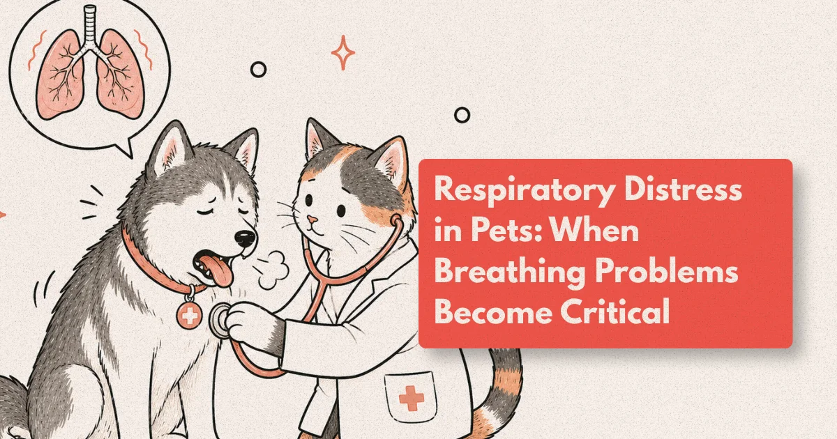
A dog panting after a walk is normal. A cat sleeping with her mouth open is not. The gap between routine respiratory variation and a life-threatening crisis can be as narrow as thirty minutes, and most owners don’t know which side they’re on until the situation has already escalated. Respiratory distress is among the most time-sensitive emergencies in veterinary medicine, accounting for approximately 15-20% of all after-hours emergency consultations (VECCS, 2024). Across dogs and cats, causes range from infections and allergies to heart disease, trauma, and cancer. Each requires a different emergency response, and a wrong assumption about the cause can cost an animal its life.
This guide covers what to look for, why cats are disproportionately at risk, how cardiac disease fits into the picture, and what modern emergency veterinary care including oxygen therapy and intensive monitoring actually involves.
What to Know
Respiratory distress accounts for roughly 1 in 6 veterinary emergency visits, and the time from symptom onset to oxygen supplementation directly affects survival odds. Cats with open-mouth breathing are in acute crisis. In dogs, blue or gray gums signal critical hypoxemia requiring immediate emergency care (VECCS, 2024).
The Signs You Need to Act On
Breathing is automatic enough that changes often go unnoticed until they become impossible to ignore. A healthy dog at rest takes 10-30 breaths per minute; a cat, 20-30. When those numbers climb above 40 at rest, or when a pet is visibly straining with each breath, something is wrong inside.
Some signs are easy to miss. Others aren’t. Blue or gray gums, where the normal pink color has been replaced by something closer to slate, mean the pet’s tissues aren’t getting enough oxygen. That’s a crisis by any clinical definition. Open-mouth breathing in a cat sits in the same category. Cats almost never breathe through their mouths. When they do, it means nasal breathing is no longer sufficient.
Other warning signs worth knowing:
- Rapid, shallow breathing at rest or during sleep
- Elbows-out posture with the neck extended (orthopnea, the body’s way of maximizing chest expansion)
- Visible abdominal pumping with each breath, where the belly contracts hard to push air out
- Audible wheezing, crackling, or a high-pitched stridor with each inhale or exhale
- Sudden reluctance to lie down
- Collapse or sudden weakness
Any one of these warrants immediate emergency care. Don’t take a wait-and-see approach with breathing.
Why Cats Are Uniquely Vulnerable to Breathing Crises
Cats are stoic by design. A dog in respiratory distress often pants, paces, and makes sounds that make the problem obvious. A cat in the same condition may go quiet, sit still in an unusual posture, and show almost nothing outwardly. By the time a cat looks visibly distressed to an average owner, the condition is typically advanced.
Feline asthma is one of the most common drivers of cat breathing problems, affecting an estimated 1-5% of cats globally, with Siamese breeds showing a disproportionately higher prevalence (Journal of Veterinary Internal Medicine, 2022). During an episode, the lower airways constrict and fill with mucus, dramatically restricting airflow. From the outside, it can look like gagging or retching. It isn’t. Cats caught in an asthmatic episode often crouch with their shoulders hunched, breathing with visible abdominal effort.
Pleural effusion, the accumulation of fluid around the lungs inside the chest cavity, is another leading cause of feline respiratory distress. A study in the Journal of Small Animal Practice found that cats presenting with pleural effusion had typically been showing subtle signs for 72 hours or more before their owners recognized distress (JSAP, 2021). The fluid compresses the lungs progressively, and cats compensate through postural adjustments and reduced activity until they can’t anymore.
A pattern seen consistently in clinical settings is that cats with moderate pleural effusion continue eating and drinking until fluid volume crosses a critical threshold. At that point, decompensation can appear sudden, even though the process has been building for days.
From clinical observation: Cats with pleural effusion are routinely underestimated by owners because the animal often sits quietly rather than showing outward agitation. The elbows-out posture and reluctance to lie on one side are the key early tells, signs that are easy to attribute to “being grumpy” rather than “struggling to breathe.”
Other conditions specific to cats include chylothorax (lymphatic fluid in the chest, often linked to cardiac or mediastinal disease), pyothorax (pus in the thoracic cavity, usually from bite wounds or bacterial spread), and diaphragmatic hernia following blunt trauma.
Open-mouth breathing in a cat is, without exception, a medical emergency until proven otherwise. It signals the point where nasal breathing is no longer sufficient. There is no “wait and see” with this presentation.
According to a 2023 study in the Journal of Feline Medicine and Surgery, nearly 40% of owners who witnessed open-mouth breathing in their cats waited more than two hours before seeking emergency care, a delay that materially worsens prognosis in conditions like pleural effusion and feline asthma (JFMS, 2023). Immediate transport to emergency care, without waiting for a regular veterinary appointment, is the correct response.
When the Heart Is the Problem
Cardiovascular disease is among the most significant causes of respiratory distress in pets, and it’s also frequently misunderstood by owners. The respiratory symptoms don’t originate primarily in the lungs. They’re the downstream effect of a heart that’s no longer pumping effectively. When the heart fails, fluid backs up. In dogs, that typically means fluid accumulating in the lung tissue itself (cardiogenic pulmonary edema). In cats, it most often means fluid collecting around the lungs (pleural effusion).
In small and medium-breed dogs, mitral valve disease (MVD) is the most common cardiac culprit. The mitral valve leaks blood backward, raising pressure in the pulmonary vasculature and ultimately pushing fluid into the lungs. Research published in the Journal of Veterinary Cardiology found that MVD affects over 50% of Cavalier King Charles Spaniels by age 5, and nearly all individuals by age 10 (Swenson et al., J Vet Cardiology, 1996; updated epidemiological data 2023). Other frequently affected small breeds include Dachshunds, Poodles, and Chihuahuas.
In large-breed dogs, dilated cardiomyopathy (DCM) is more common. The heart muscle weakens, chambers enlarge, and pumping efficiency drops. Doberman Pinschers carry a particularly heavy burden: a European multicenter study found a lifetime DCM risk exceeding 58% in the breed (Wess et al., J Vet Intern Med, 2010).
For cats, the dominant cardiac disease is hypertrophic cardiomyopathy (HCM), where the left ventricular wall thickens and the heart’s filling capacity decreases. Maine Coons and Ragdolls carry known genetic mutations (MYBPC3 variants) that elevate HCM risk significantly. Prevalence in the general cat population is estimated at 15% (JFMS, 2022), making HCM the single most common heart disease in cats, and one of the leading reasons cats arrive in respiratory crisis with no prior clinical history.
The clinical distinction between cardiac and non-cardiac respiratory distress matters because the treatments differ fundamentally. A pet in heart failure given bronchodilators for presumed asthma may deteriorate rather than improve. The definitive work-up includes chest radiographs to assess cardiac silhouette and fluid distribution, echocardiography to evaluate chamber dimensions and valve function, an ECG for arrhythmia detection, and blood pressure measurement. For established cases, referral to a board-certified veterinary cardiologist offers the most precise treatment optimization, particularly around timing and titration of medications like furosemide, pimobendan, and ACE inhibitors.
A pattern worth noting clinically: Cats with HCM frequently present with acute respiratory distress as the first detected sign of heart disease. The underlying cardiac remodeling may have been progressing silently for months. This makes HCM cats difficult to catch before the crisis, and is why any middle-aged or senior cat with sudden breathing difficulty warrants a cardiac work-up even in the absence of a known cardiac history.
Oxygen Therapy: The Life-Saving First Intervention
Before a diagnosis is confirmed, oxygen is already helping. Supplemental oxygen is the universal first response to respiratory distress in veterinary medicine. It raises blood oxygen saturation (SpO2), buys time for diagnostic work, and reduces the metabolic stress of the patient working against hypoxemia. Almost everything else can wait until the patient is breathing comfortably. Oxygen therapy can’t.
Veterinary oxygen delivery comes in several forms, and the choice depends on the severity of distress and patient temperament.
Flow-by oxygen is the simplest approach: a tube or mask held near the nose, providing passive enrichment without restraint. It works for mildly distressed patients who’d be further stressed by physical handling. The concentration is imprecise, but it’s meaningfully better than room air.
Oxygen cage therapy is the gold standard for severely distressed or fractious patients, particularly cats. A sealed chamber maintains oxygen concentrations of 40-60%, compared to 21% in ambient air. The pet breathes freely without being touched, which matters enormously for cats, where handling-related stress can trigger fatal decompensation. A well-managed oxygen cage can restore SpO2 above 95% within 15-20 minutes in most patients presenting with moderate respiratory distress (Plumb’s Veterinary Drug Handbook, 2023).
High-flow nasal oxygen (HFNO) delivers humidified oxygen through a small catheter placed in one or both nostrils, providing higher and more consistent concentrations than flow-by. It’s better tolerated by calm dogs and is increasingly standard in veterinary intensive care. In human critical care, HFNO reduces the need for intubation by 30-40% in hypoxemic patients; veterinary pilot studies are showing similar trends (Vet Critical Care, 2024).
Mechanical ventilation is reserved for patients in acute respiratory failure who can’t maintain acceptable oxygenation through supplemental methods alone, or those requiring thoracic surgery under anesthesia. This capability is available at specialist referral centers and veterinary academic hospitals.
Pulse oximetry guides every decision. An SpO2 reading below 94% at rest is the threshold for active oxygen intervention in most veterinary protocols. Below 90% is critical. The clinical goal is to hold saturation above 95% while the team identifies and addresses the underlying cause.
A resting SpO2 below 94% is the widely accepted threshold for oxygen supplementation in small animals, and below 90% constitutes a critical emergency requiring immediate escalation beyond flow-by delivery (Plumb’s Veterinary Drug Handbook, 2023). Early intervention consistently improves patient stability before diagnosis is confirmed, buying the time needed for chest radiographs, ultrasound, and bloodwork.
Inside the Veterinary ICU: Intensive Monitoring and Intervention
When oxygen therapy alone doesn’t stabilize a patient, or when the case is complex enough to require frequent reassessment and rapid treatment adjustments, the pet moves to intensive care. Veterinary ICUs operate on the same principles as human critical care: continuous monitoring, tight treatment titration, and a team positioned to respond to any change in status immediately.
A pet admitted for respiratory distress will typically receive continuous pulse oximetry, with the probe attached to the tongue, inner ear, or toe web. An IV catheter goes in for medication delivery without repeated handling. Fluid therapy is one of the most carefully calibrated interventions in the ICU: too much worsens cardiogenic pulmonary edema; too little in a shock state compromises cardiac output. Getting that balance right is a large part of what intensive care management involves.
What has changed emergency veterinary medicine significantly in recent years is point-of-care ultrasound (POCUS). A skilled clinician can now identify pleural effusion, pericardial effusion, or pulmonary edema at the bedside in under three minutes (Journal of Veterinary Emergency and Critical Care, 2023). That speed changes how quickly targeted treatment can begin. In a deteriorating patient, three minutes is not abstract.
Serial chest radiographs and blood gas analysis track whether the patient is responding. Specific interventions follow the diagnosis: thoracocentesis to drain pleural effusion (which often provides near-immediate respiratory relief), bronchodilators for asthma, IV furosemide for congestive heart failure, or broad-spectrum antimicrobials for pyothorax.
ICU survival rates reflect the range of conditions presenting with respiratory distress. Pets with infectious pneumonia treated aggressively with appropriate antibiotics recover in 70-80% of cases. Those in acute congestive heart failure stabilized within the first four hours have a survival-to-discharge rate of approximately 60-75%, though long-term prognosis depends on the underlying cardiac diagnosis (VECCS Annual Reports, 2023).
Owners are often surprised that ICU visitation is restricted. It isn’t a bureaucratic policy. A pet that sees its owner will attempt to reach them. In a hypoxic animal, that exertion can trigger decompensation. The restriction is physiological protection, not a barrier.
What to Do Before You Get to the Clinic
Home response is not treatment. Respiratory distress can’t be reversed without veterinary support. But you can significantly affect the outcome by acting correctly and moving quickly.
Stay calm. Pets mirror their owner’s anxiety, and an elevated stress response increases oxygen demand in an animal that’s already struggling to maintain it.
Don’t restrict the pet’s movement or position. Let it find whatever posture makes breathing easiest. Cats in distress often stay upright; forcing one to lie flat can accelerate decompensation. Dogs may want to stand with their elbows wide. Let them.
Don’t muzzle a dog that’s struggling to breathe. Muzzles restrict airflow substantially. In a dog that’s already hypoxic, that restriction can be fatal within minutes.
Call ahead to the emergency clinic while you’re already in the car. “My cat is breathing rapidly with effort, we’re 10 minutes away” triggers preparation. An oxygen cage will be ready at intake rather than five minutes after your arrival. That gap is not trivial when the window is narrow.
Frequently Asked Questions
How can I tell if my cat’s breathing is abnormal at rest?
Count breaths for one full minute while your cat is sleeping or completely relaxed. A resting respiratory rate above 30 breaths per minute is abnormal and warrants same-day veterinary contact. Rates above 40, open-mouth breathing, or visible abdominal effort at any respiratory rate are emergencies (JFMS, 2022).
What does oxygen therapy for pets actually involve at the clinic?
Most clinics start with an oxygen cage, a sealed unit maintaining 40-60% oxygen concentration versus 21% in room air. Your pet breathes freely in the enriched environment without restraint. Nasal cannulas or masks may follow once the patient is stable. A well-managed oxygen cage restores SpO2 above 95% within 15-20 minutes in most moderate cases (Plumb’s Veterinary Drug Handbook, 2023).
Can heart disease cause sudden respiratory distress with no prior warning?
Yes, particularly in cats with HCM. The cardiac remodeling progresses silently over months, and pleural effusion accumulates gradually until a threshold is crossed. Many cats present in acute distress with no prior clinical history. An echocardiogram is the only reliable way to detect HCM before the crisis point (JFMS, 2022).
What happens during respiratory stabilization in a veterinary ICU?
Continuous pulse oximetry, IV medication access, serial chest imaging, and targeted interventions like thoracocentesis or bronchodilator delivery are the core elements. Point-of-care ultrasound now allows bedside diagnosis of chest fluid or edema in under three minutes, enabling treatment decisions without waiting for a radiograph (JVECCS, 2023).
Are brachycephalic breeds at higher risk for respiratory emergencies?
Yes. Bulldogs, French Bulldogs, Pugs, and Persian cats have structural airway narrowing that creates chronic respiratory compromise, worsens in heat, and amplifies any secondary condition. Over 50% of brachycephalic dogs have measurable airway obstruction even at rest (BSAVA, 2023). These breeds benefit from proactive surgical evaluation before an emergency arises.
The Window That Matters
Respiratory distress doesn’t give much time for reflection. What it gives is a narrow window, sometimes hours, sometimes less, where the right response changes everything. Oxygen buys that window. ICU monitoring uses it productively. Cardiology, targeted treatment, and careful diagnosis close it on favorable terms.
None of that sequence starts without recognizing the signs early. Open-mouth breathing in a cat, blue gums in a dog, an unusual resting posture, or simply a pet that won’t lie down: these aren’t things to observe overnight. They’re reasons to move.
The pets that survive respiratory crises aren’t always the ones with mild conditions. They’re most often the ones whose owners acted before the window closed.