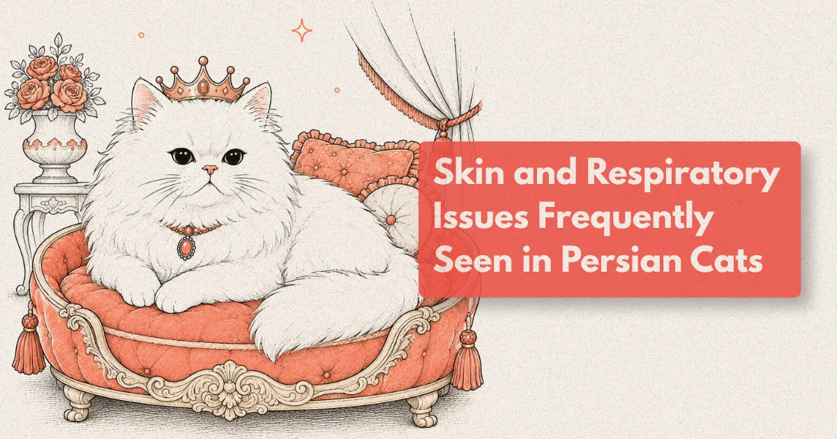
The Persian cat is one of the oldest and most widely kept pedigree breeds in the world, and it brings to that status a set of anatomical characteristics that have direct and consistent clinical consequences. The extremely foreshortened skull, compressed nasal structure, abundant facial skin folds, and dense long coat that define the breed’s appearance also define the health challenges that practitioners working with Persians encounter in routine clinical practice. Understanding those challenges as a connected system, rather than as isolated problems, is the basis of effective preventive and therapeutic care for the breed.
Two clinical categories account for the majority of Persian-specific health presentations: brachycephalic respiratory syndrome and skin fold disease. They are linked not only by the same underlying anatomy but by a clinical pattern in which both are underrecognised in their early stages, both progress if unmanaged, and both have substantially better outcomes when identified and addressed before secondary disease has established. The coat adds a third dimension, since coat condition in a Persian is simultaneously an aesthetic concern, a welfare indicator, and a clinical sign.
What to Know
Approximately 80% of Persian and Exotic Shorthair cats examined in a structured clinical study showed some form of respiratory sign at rest, including stertor, stridor, or increased respiratory effort (Farnworth et al., PLOS ONE, 2016). Persian cat breathing issues attributable to brachycephalic obstructive airway syndrome are diagnosable and in many cases surgically correctable, yet most affected cats reach adulthood without a formal respiratory diagnostics assessment.
The Persian Face: Anatomy With Clinical Consequences
The defining feature of the Persian skull is an extreme reduction in the nasofacial ratio: the distance between the nose tip and the inner corner of the eye is proportionally far smaller than in the domestic shorthaired cat. This compression is not limited to external appearance. The nasal passages, nasopharynx, and associated soft tissue structures are all proportionally reduced or distorted, with consequences for airflow, drainage, and the functional capacity of the upper respiratory tract under normal resting conditions.
The nasal fold, the skin ridge that runs across the bridge of the nose and continues into the periorbital and perinasal regions, is prominent in most Persians and absent or minimal in the mesocephalic cat. Its clinical relevance is that it creates a warm, moist, friction-prone microenvironment in direct contact with the medial canthus and nasolacrimal drainage system. Epiphora, the overflow of tears onto the facial skin, is almost universal in the breed. Where tears chronically contact the fold, secondary bacterial and Malassezia colonisation and dermatitis develop with predictable regularity.
Polycystic kidney disease (PKD), caused by a dominant mutation in the PKD1 gene, is the most significant systemic inherited condition in the breed. Lyons and colleagues demonstrated prevalence of 36-49% in unscreened Persian populations (2004). PKD is not a respiratory or dermatological condition, but it is part of the complete breed health picture and should be screened for by DNA testing or ultrasound in all Persians presented for any wellness assessment.
The coat of the Persian, while not a disease in itself, requires daily professional-standard grooming to remain free of matts, debris accumulation, and the secondary skin irritation that persistent matting produces. In older, painful, or unwell Persians, grooming behaviour declines and coat condition deteriorates as a consequence. This means coat condition is a clinical signal: a Persian presented with a significantly degraded coat, in the absence of any owner-reported change in grooming routine, deserves a full clinical assessment for any underlying condition that may be reducing the cat’s capacity or willingness to self-groom.
Persian Cat Breathing Issues: Recognising BOAS in Cats
Brachycephalic obstructive airway syndrome (BOAS) in cats encompasses the anatomical abnormalities that collectively reduce airflow through the upper respiratory tract. In Persians, the primary components are stenotic nares (narrowed nostril openings that increase inspiratory resistance), aberrant intranasal turbinates (rostral nasal turbinate tissue that extends abnormally into the nasal passage and further obstructs airflow), an elongated and thickened soft palate, and a relatively narrowed nasopharynx.
Farnworth and colleagues (PLOS ONE, 2016) assessed 50 brachycephalic cats and found that the majority demonstrated respiratory signs at rest, including stertor (a low-pitched snoring noise produced by soft tissue vibration in the pharynx), stridor (a higher-pitched noise indicative of narrowed upper airway segments), and open-mouth breathing during mild handling or minimal exertion. These signs were present in cats whose owners reported them as normal and whose clinical records contained no respiratory concern.
This owner normalisation is the core clinical problem. An owner who has kept Persians for years, or who grew up with the breed, often has a calibration of normal that accommodates significant respiratory compromise. The cat that snores continuously, breathes audibly at rest, sleeps with its mouth slightly open, and avoids exercise is not normal: it is a cat with chronic upper airway obstruction that has adapted its lifestyle to reduce the respiratory demand it cannot comfortably meet.
The clinical grading of feline BOAS follows a spectrum. Mild cases show audible stertor at rest without measurable exercise intolerance. Moderate cases show significant stertor with an elevated respiratory rate, exercise intolerance, and at least occasional open-mouth breathing. Severe cases show constant open-mouth breathing, cyanosis, or syncopal episodes, and represent acute welfare concerns requiring urgent assessment and intervention.
Sleep quality is a clinically underexplored dimension of feline BOAS. In dogs, BOAS severity correlates with documented sleep disturbance and daytime somnolence. The same relationship is likely in cats, and the owner observation that a Persian sleeps excessively, sleeps in unusual positions (sternal or with head elevated to open the airway), or is persistently fatigued is clinically meaningful.
Respiratory Diagnostics: Assessing the Upper Airway
The respiratory diagnostics pathway for a Persian cat with suspected BOAS begins with a structured physical examination under minimal stress, since handling alone can precipitate acute respiratory compromise in a severely affected cat. Observation before handling, noting respiratory rate, rhythm, nostril excursion, and the presence of audible respiratory noise at rest, provides baseline information that direct examination may obscure.
Rhinoscopy, performed under general anaesthesia, allows direct visualisation of the nasal cavity and assessment of turbinate anatomy, mucosal health, and the presence of polyps, discharge, or structural abnormality. It is indicated in Persian cats with chronic nasal discharge, sneezing, or unilateral respiratory signs that may have infectious, inflammatory, or structural causes beyond straightforward BOAS.
Computed tomography (CT) is the imaging modality that most completely characterises the upper airway anatomy in brachycephalic cats. CT evaluates the degree of nasal turbinate intrusion, the nasopharyngeal cross-sectional area, soft palate thickness and length, and any concurrent middle ear changes (brachycephalic cats have higher rates of middle ear effusion and primary secretory otitis media). For surgical planning, CT is invaluable: knowing the three-dimensional anatomy of the obstruction before operating determines which procedures are indicated and in what combination.
Thoracic radiography is part of the baseline evaluation for any Persian with significant respiratory signs. It assesses for cardiomegaly, pleural effusion, pulmonary infiltrate, and tracheal diameter. Brachycephalic cats have a higher prevalence of tracheal hypoplasia (a congenitally narrowed trachea) than mesocephalic cats, and this finding changes both the prognosis for upper airway surgery and the clinical management threshold.
Bronchoalveolar lavage (BAL) is indicated when lower respiratory tract disease is suspected, providing cytology and culture samples that characterise infection, inflammation, or neoplastic processes that upper airway assessment cannot reach.
Facial Fold Dermatitis: The Most Consistently Underestimated Problem
Facial fold dermatitis in Persian cats is the skin condition that owners most commonly accept as a permanent, unalterable feature of the breed rather than a diagnosable and manageable clinical problem. The premise is understandable: the skin fold is permanent, so the associated redness and discharge seem to follow. The clinical reality is that the severity of fold dermatitis is highly variable and directly influenced by management, that secondary infection significantly worsens welfare beyond what the fold alone produces, and that appropriate treatment substantially reduces both clinical signs and the cat’s daily discomfort.
The nasal fold creates a pocket of warm, humid skin in permanent contact with episcleral and nasolacrimal secretions. The combination of moisture, warmth, reduced air circulation, and organic material provides an ideal environment for colonisation by Staphylococcus spp., Malassezia pachydermatis, and gram-negative bacteria. The erythema, malodour, brown discharge, and skin thickening that characterise established fold dermatitis in Persians are not caused by the fold anatomy itself but by the secondary microbial populations it supports.
Clinical assessment of fold dermatitis should include cytology of the fold material, distinguishing between simple maceration, bacterial pyoderma, and yeast dermatitis, since the topical treatment differs meaningfully across these categories. Bacterial fold pyoderma responds to topical antiseptic or antibiotic preparations; Malassezia fold dermatitis requires antifungal treatment; mixed infections require both. Attempting to manage fold dermatitis without cytological direction is the reason many treatment attempts fail.
Epiphora management is inseparable from fold dermatitis management. The nasolacrimal duct in Persian cats is frequently non-functional or partially obstructed due to the compression of the duct by the foreshortened skull anatomy. Where the duct functions normally, flushing it periodically keeps the drainage pathway clear. Where obstruction is structural and permanent, the epiphora is managed rather than resolved: daily gentle cleaning of the medial canthus and fold with an appropriate veterinary wipe, maintaining a dry surface, and monitoring for secondary infection.
From clinical practice: Persian cats presented for the first time in mature adulthood frequently have fold dermatitis that has been present and worsening for years without treatment, because neither the owner nor previous practitioners had reframed it as a treatable condition rather than a breed characteristic. The cat with a chronic brown-stained nasal fold that has been present since kittenhood is not a cat where treatment “won’t help.” It is a cat where appropriate fold management, introduced consistently, typically produces visible improvement within three to four weeks, and where the owner, on seeing that improvement, understands for the first time that the cat had been experiencing daily discomfort that was being caused and maintained by inadequate management rather than by the anatomy alone.
Dermatophytosis: A Persian-Specific Clinical and Public Health Concern
Dermatophytosis (ringworm, caused primarily by Microsporum canis in cats) has a disproportionate clinical significance in Persians for two reasons that are often conflated but should be held separately. The first is that Persians are substantially over-represented among cats with active clinical dermatophytosis: their long, dense coat provides an environment that supports fungal growth, and any skin fold irritation or coat damage creates entry points for infection. The second is that Persian cats are recognised as disproportionately common asymptomatic or minimally symptomatic carriers of M. canis, shedding viable spores in scale and hair without developing obvious lesions.
The public health implication of the carrier issue is significant and underappreciated in the ownership conversation. A household with a Persian cat and young children, elderly individuals, or immunocompromised family members has a meaningful dermatophyte exposure risk even if the cat appears clinically normal. The WSAVA/World Association for Veterinary Dermatology consensus guidelines (Moriello et al., Veterinary Dermatology, 2017) recommend that cats from high-prevalence populations, including Persians in multi-cat environments and catteries, undergo routine fungal culture screening even without clinical signs.
Treatment of confirmed dermatophytosis in Persians requires both topical and systemic antifungal therapy and environmental decontamination. The dense coat of a Persian is challenging to treat topically because achieving adequate contact between antifungal solution and skin surface requires either clipping or very thorough application. Systemic itraconazole or terbinafine, combined with twice-weekly antifungal shampoo treatment, is the evidence-based approach for feline dermatophytosis (Moriello et al., 2017). Treatment duration should be guided by culture negativity, not a fixed number of weeks: treatment should continue until two consecutive fungal cultures taken two to three weeks apart are negative.
Coat Condition as a Clinical Indicator
The Persian coat is both a management requirement and a diagnostic resource. An owner who maintains a Persian’s coat correctly attends to it daily and has an intimate familiarity with its normal texture, the areas that matt first, the rate of growth, and the baseline coat density. Changes in any of these are clinically detectable before other signs of systemic illness, and the informed Persian owner is a valuable source of early clinical information.
Progressive matt formation in a previously well-maintained Persian coat, occurring despite no change in grooming routine, suggests either that the coat is changing (altered oil production, texture change suggesting endocrine or nutritional change) or that the cat is resisting grooming in a way it previously did not. Both warrant clinical assessment. A cat that is flinching or withdrawing from grooming contact over the dorsum or hindquarters is a cat with possible musculoskeletal pain. A cat whose coat is producing more matts than usual despite similar management may have seborrhoea, a nutritional imbalance, or an underlying systemic condition affecting coat quality.
Primary idiopathic seborrhoea is recognised as a heritable condition in Persians, producing excessive scaling and greasiness of the coat from a young age. Secondary seborrhoea, developing in a previously normal coat, is a dermatological sign requiring investigation. Diagnosis involves skin cytology, fungal culture, and assessment for the underlying causes of secondary seborrhoea including ectoparasites, allergic skin disease, nutritional deficiency, and systemic illness.
Omega-3 fatty acid supplementation at appropriate therapeutic doses supports skin barrier function and reduces inflammatory mediator production in the skin. For Persian cats with mild to moderate coat condition issues without confirmed systemic cause, dietary supplementation with fish oil providing EPA and DHA is a reasonable adjunct while further investigation is ongoing.
Building the Long-Term Care Plan for Persian Cats
The Persian cat’s health management programme is best understood as an integration of several ongoing requirements rather than a series of isolated presentations. Respiratory function needs assessment from kittenhood, not at the point when the owner notices that breathing has become laboured. Skin fold management is a daily home-care practice, not a treatment initiated when dermatitis has become infected. Coat care is a preventive health measure, not an aesthetic preference. PKD screening is a one-time genetic test that establishes whether the kidney disease trajectory needs to be monitored.
Surgical management of BOAS in Persian cats has the same principle as in brachycephalic dogs: remove or reduce the anatomical obstructions that are impairing airflow before the compensatory increased respiratory effort and chronic airway inflammation cause secondary changes. Stenotic nares correction is the most accessible and frequently most beneficial procedure. In experienced hands, it can be performed under brief general anaesthesia and produces immediate reduction in inspiratory resistance. Soft palate shortening and intranasal turbinectomy are performed in more severely affected cats where nares correction alone is insufficient.
Post-surgical outcomes for feline BOAS correction are positive in the majority of cases when surgery is performed on appropriately selected patients before severe secondary laryngeal changes develop. The evidence base is smaller than for canine BOAS correction, but the clinical experience reported in specialist practice is that owner-reported quality-of-life improvement is significant, with reduced respiratory noise, improved exercise tolerance, and better sleep quality documented.
The practical message for Persian cat owners is that breed-specific monitoring begins with the first wellness visit and does not end. Respiratory function, skin fold condition, coat health, body weight, blood pressure, and renal function screening are all components of a programme that, maintained consistently, gives the majority of Persians the best possible chance of a healthy, comfortable life within the anatomical constraints their breeding has created.
Frequently Asked Questions
Do all Persian cats have breathing problems?
The anatomical changes associated with Persian brachycephaly are present to some degree in virtually all individuals of the breed. Whether those changes produce clinically significant breathing problems depends on severity. Studies suggest the majority of Persians have some degree of respiratory sign at rest, but many owners do not recognise these signs as abnormal. A structured respiratory assessment by a veterinarian familiar with brachycephalic feline medicine provides the most accurate individual evaluation.
What does facial fold dermatitis look like in a Persian cat?
Early facial fold dermatitis presents as redness and mild moisture in the skin fold along the nasal bridge and adjacent to the inner corners of the eyes. As it progresses, there is visible brown discharge, an unpleasant odour, and thickening of the fold skin. Secondary bacterial or yeast infection is common in established cases. Treatment requires cytology to identify the microbial populations involved and targeted topical therapy. Daily home fold cleaning with a veterinary-approved product prevents and controls mild cases.
Can Persian cat breathing problems be treated surgically?
Yes. Stenotic nares correction is the most commonly performed and effective procedure, widening the nostril openings to reduce inspiratory resistance. Soft palate shortening and intranasal turbinectomy are performed in more severely affected cats. These procedures are best undertaken before secondary laryngeal changes develop from years of compensatory increased respiratory effort. Results are generally positive, with most owners reporting significant improvement in respiratory noise, activity tolerance, and sleep quality.
How do I know if my Persian's coat condition indicates a health problem?
Any progressive deterioration in coat condition without a corresponding change in grooming routine warrants assessment. Watch for new matt formation in unusual locations, increased scale or greasiness, hair loss, or the cat resisting grooming. Normal coat changes with ageing are gradual and diffuse; sudden or regionally specific changes are more likely to indicate a medical cause. A basic skin examination including cytology, fungal culture, and parasitology screen is the appropriate first investigation.
What is PKD and should my Persian cat be tested?
Polycystic kidney disease is a hereditary condition affecting approximately one third to one half of unscreened Persians, caused by a dominant gene mutation that produces progressive kidney cyst formation. DNA testing identifies the mutation from a simple cheek swab. Cats testing negative are confirmed non-carriers. Cats testing positive benefit from regular ultrasound monitoring, early nutritional management, and blood pressure surveillance. PKD testing should be performed once in every Persian and Himalayan cat regardless of clinical signs.