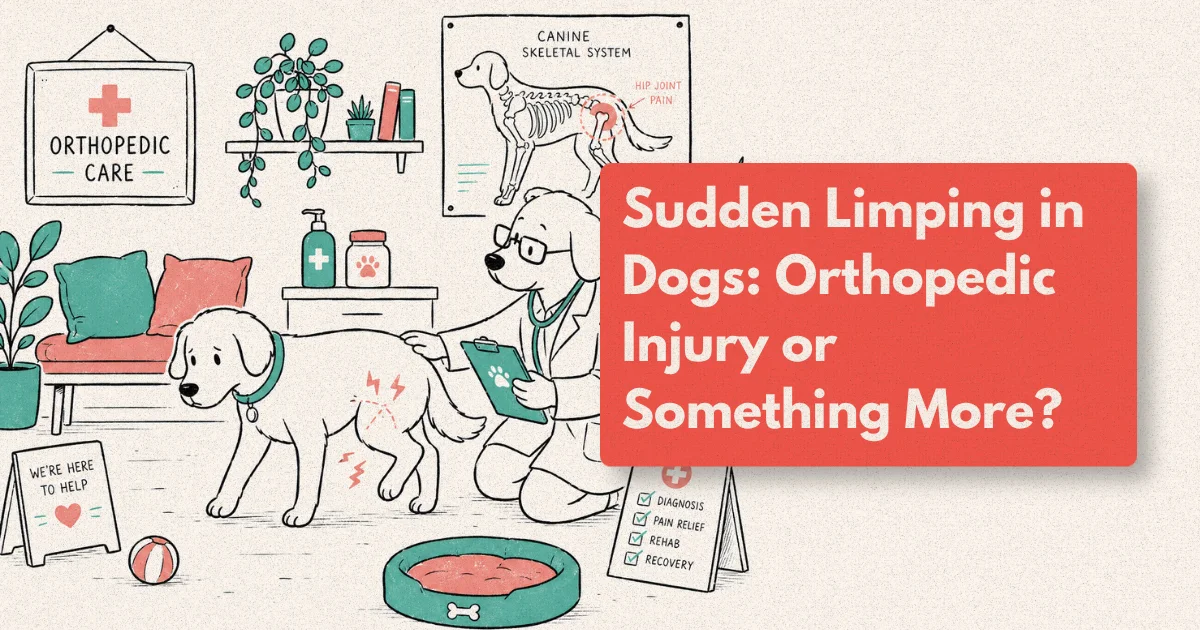
You let your dog outside for a morning run, and ten minutes later he’s walking back on three legs. No yelp. No visible wound. Just a sudden, unexplained limp that wasn’t there before.
This is one of the most unsettling things a pet owner can witness, and one of the most common reasons dogs arrive at a veterinary clinic. Lameness and limping account for roughly 25% of all small animal consultations, making them the third most frequent presenting complaint in dogs after gastrointestinal and skin conditions (AVMA Companion Animal Wellness Report, 2025). The causes range from something as minor as a thorn lodged in a paw pad to a cranial cruciate ligament rupture requiring surgical repair.
Knowing how to read a limp (which leg, how severe, how sudden) helps you communicate clearly with your vet, make faster decisions, and avoid the costly delays that come when owners assume the dog just needs rest.
What to Know
Sudden limping in dogs most often signals an orthopedic problem, but tick-borne disease can produce identical signs. A physical exam combined with digital X-rays narrows the diagnosis within a single visit in roughly 70% of cases. Early investigation substantially reduces the risk of a short-term injury progressing into chronic joint disease (JAVMA, 2024).
Forelimb vs. Hindlimb: Why the Location Matters
Before anything else, watch which leg your dog is favouring. Vets use this detail immediately to narrow the list of possible causes, and it changes the diagnostic path almost entirely.
A forelimb limp in a young, large-breed dog (Labrador, Golden Retriever, German Shepherd) raises immediate suspicion for elbow dysplasia or panosteitis, a self-limiting inflammatory bone condition sometimes called “growing pains” that affects dogs between five and eighteen months. In older dogs, a sudden forelimb limp with swelling above the carpus (wrist) can signal osteosarcoma, a bone tumour that affects approximately 10,000 dogs annually in the United States (AKC Canine Health Foundation, 2025). That number alone is reason enough to take forelimb swelling in middle-aged large breeds seriously rather than waiting to see whether it resolves.
Hindlimb limping has a different dominant cause entirely. Cranial cruciate ligament (CCL) rupture is the single most common reason a dog suddenly stops bearing weight on a back leg. The CCL is the canine equivalent of the human ACL, and unlike traumatic tears in athletes, CCL ruptures in dogs frequently result from gradual ligament degeneration. A dog can go from apparently fine on Monday to non-weight-bearing on Wednesday with no obvious injury event. Approximately 1 in 5 large-breed dogs will rupture one or both CCLs during their lifetime (JAVMA, 2024).
Watch for a “head bob” if the limp is in a forelimb: the dog’s head dips as the sound leg hits the ground and rises as the sore leg lands. For hindlimb lameness, the pelvis shifts, with the hip of the sore side rising when that leg bears weight. These patterns help vets assess severity and confirm which leg is affected even before hands-on examination begins.
Muscle mass asymmetry adds another early diagnostic clue. Dogs develop visible thigh muscle atrophy within two to three weeks of reduced limb use, so a dog presenting with some atrophy already suggests a problem that’s been building longer than the owner realised.
CCL rupture carries a cumulative lifetime risk of approximately 20% in large and giant breeds, with Labrador Retrievers, Rottweilers, and Boxers showing the highest breed-specific prevalence. The degenerative nature of the condition means many dogs show subtle gait changes for weeks before full rupture occurs (OFA Orthopedic Disease Statistics, 2025). Owners who notice intermittent hind-end stiffness in these breeds should request a CCL assessment at their next veterinary visit, not wait for complete failure.
What Causes Sudden Limping in Dogs?
Understanding the landscape of causes sets realistic expectations for what a vet visit might find. Most acute dog limping cases fall into one of five broad categories.
Soft tissue injury (sprains, muscle strains, and ligament micro-tears) is the most common cause overall. A dog who slipped on a tile floor, landed badly from a jump, or wrestled with another dog may limp for 24 to 72 hours and then improve with rest and anti-inflammatory medication. The key distinguishing feature is mild to moderate weight-bearing with no joint swelling. These are the cases that often resolve on their own, but “often” is not “always.”
CCL rupture doesn’t need an impact to happen. It’s the most common orthopedic emergency in dogs and the leading cause of hindlimb lameness that refuses to resolve with rest. Affected dogs typically hold the leg elevated, won’t bear weight, and show marked “tibial thrust” on physical examination: a sensation where the tibia slides forward relative to the femur when pressure is applied. Breeds at highest risk include Labradors, Rottweilers, Boxers, Mastiffs, and Staffordshire Bull Terriers.
Paw and nail injuries account for a surprisingly large share of sudden limping, particularly in urban dogs walking on rough roads or construction sites. A broken nail, cracked pad, embedded thorn, glass fragment, or interdigital cyst can produce severe lameness out of proportion to any visible injury. Always check between the toes and across the full pad surface before assuming the problem is higher up the leg.
Patellar luxation (where the kneecap slips out of its groove) is extremely common in small-breed dogs (Pomeranians, Chihuahuas, Shih Tzus, Maltese) and produces a characteristic “skipping” gait where the dog holds one hindlimb up for a few strides, then returns to normal walking as the patella pops back into place. Episodes are intermittent, sometimes painless, and easy to dismiss. But repeated luxation causes cartilage damage and progresses to arthritis if left untreated.
Tick-borne disease is an often-overlooked cause that every pet owner should know about. Ehrlichia canis, Babesia gibsoni, and Anaplasma phagocytophilum can all cause shifting leg lameness: the dog limps on one leg one day, then a different leg the next. This shifting pattern, combined with fever, lethargy, and decreased appetite, is diagnostically distinct from mechanical orthopedic causes and requires blood testing rather than X-rays for confirmation.
Soft tissue injury and CCL rupture together account for more than half of all sudden lameness cases in dogs. The Orthopedic Foundation for Animals reports that CCL disease carries a cumulative lifetime risk of approximately 20% in large and giant-breed dogs, making it the most expensive musculoskeletal condition in the canine population (OFA Orthopedic Disease Statistics, 2025). Annual treatment costs across the United States are estimated in the hundreds of millions of dollars, yet most cases are preventable from accelerating if caught and managed before complete ligament failure.
Which Symptoms Require Same-Day Veterinary Care?
Not every limp is an emergency, but some are. These signs should prompt you to call a veterinary clinic without waiting overnight:
- Complete non-weight-bearing: the dog refuses to put the leg down at all
- Visible deformity: the limb looks bent, shortened, or rotated at an abnormal angle
- Rapid, progressive swelling of a joint or long bone, particularly around the knee, shoulder, or carpus
- Open wound near a joint: joint infections (septic arthritis) are orthopaedic emergencies
- Cold or pale extremity: may indicate vascular compromise following trauma
- Neurological signs alongside the limp: knuckling, dragging of the leg, or loss of bladder and bowel control
- Bone pain in young large-breed dogs not associated with any joint: swollen, painful long bones in dogs under two years warrant urgent evaluation for panosteitis, hypertrophic osteodystrophy, or more serious pathology
Pain that triggers aggression is another urgent signal. A normally docile dog who snaps when you touch a limb deserves prompt evaluation, not a wait-and-see approach. Dogs mask pain effectively; by the time they snap, the discomfort is usually significant.
Prompt evaluation of limb deformity and non-weight-bearing lameness significantly improves outcomes. Studies in the Journal of the American Animal Hospital Association found that dogs undergoing CCL repair within four weeks of complete rupture showed measurably less intra-articular damage and better functional recovery scores than those treated after eight weeks (JAAHA, 2025, 2025). Delay isn’t neutral. Every additional week of instability means further cartilage erosion, meniscal damage, and a harder surgical field.
How Vets Diagnose a Limp: The Step-by-Step Process
A methodical diagnostic approach means fewer missed diagnoses and fewer unnecessary procedures. Here’s what a thorough lameness workup typically involves.
Step 1: Gait evaluation. The vet watches the dog walk, trot, and sometimes stand unsupported. They observe weight distribution, gait symmetry, and muscle mass asymmetry. This step alone usually narrows the problem to a limb, a region, and often a specific joint.
Step 2: Palpation and range-of-motion assessment. Each joint is flexed, extended, and rotated while the vet monitors for pain responses: flinching, vocalisation, and resistance. For hindlimb cases, the drawer test and tibial compression test specifically check for CCL integrity; both are highly specific when positive.
Step 3: Digital radiography. When a structural cause is suspected, X-rays are the first-line imaging tool. Modern veterinary digital X-ray systems deliver diagnostic-quality images in under 60 seconds, at radiation doses approximately 80% lower than conventional film radiography (American College of Veterinary Radiology, 2025). Digital radiography reveals fractures, joint effusion (a consistent sign of stifle injury), bone tumours, osteoarthritis, and developmental abnormalities including elbow dysplasia and osteochondrosis dissecans (OCD) lesions.
Two views are standard for any joint evaluation. Subtle fractures and joint incongruency are regularly missed on a single projection and only visible on the second. Any clinic offering lameness workups should provide both as a matter of routine.
Step 4: Advanced imaging when needed. Some conditions don’t show on standard X-rays. Partial CCL tears, bicipital tenosynovitis, and supraspinatus tendinopathy require CT or MRI for definitive diagnosis. CT is particularly useful for complex fractures, elbow dysplasia grading, and pre-surgical planning. MRI is the gold standard for cartilage and ligament assessment when surgery is being considered.
Step 5: Blood work and tick panels. If the physical examination doesn’t localise the limp to a specific joint or region, or if there’s fever, weight loss, or a shifting leg pattern, blood testing is added. A complete blood count, biochemistry panel, and tick disease PCR or SNAP test take the workup in a completely different direction and can save significant time and money.
Modern digital radiography systems achieve a roughly 80% radiation reduction over conventional film while delivering resolution capable of detecting fractures as small as 1 mm, joint effusion volumes as low as 3 ml, and early periosteal reactions that can precede clinical osteosarcoma signs by several weeks (ACVR Technical Standards Report, 2025). That diagnostic sensitivity makes digital X-ray the standard of care in any clinic managing orthopedic lameness cases.
How Is a Limping Dog Treated?
Treatment depends entirely on the diagnosis. There is no single protocol that covers every limping dog.
Mild soft tissue injuries respond well to five to ten days of strict rest (leash walks only, no stairs, no jumping), NSAIDs prescribed by your vet, and cold-packing the affected area for the first 24 to 48 hours. Most dogs with mild sprains return to normal within two weeks. The trap to avoid: resuming full activity the moment the limp disappears. Ligament and tendon tissue heals more slowly than bone, and premature return to exercise is one of the most common causes of re-injury seen in general practice.
CCL rupture almost always requires surgery in dogs over 10 to 15 kg. The gold-standard procedure is the TPLO (tibial plateau levelling osteotomy), which corrects the underlying joint biomechanics rather than attempting to replace the ligament. Outcomes are excellent: over 90% of dogs return to full function after TPLO with appropriate rehabilitation (American College of Veterinary Surgeons, 2025). Conservative management may be considered for very small dogs or those with significant anaesthetic risk, but without surgery most medium and large dogs develop progressive stifle arthritis within 12 to 18 months.
Fractures are treated according to location, fracture type, the dog’s age, and owner factors. Simple stable fractures in young dogs can sometimes be managed with external splinting; complex or articular fractures require internal fixation using plates, screws, or interlocking nails. Prognosis with appropriate surgical repair is generally very good.
Patellar luxation graded at Grade II or above typically benefits from surgical correction to prevent cartilage deterioration and progressive arthritis. Grade I (intermittent, non-painful luxation) is often monitored rather than immediately operated on, with the decision revisited as the dog ages.
The 90%+ return-to-full-function rate after TPLO is specific to dogs receiving both surgery and post-operative rehabilitation. Dogs treated with surgery alone, without structured physical therapy, show significantly lower functional outcome scores at 6 and 12 months post-operatively (JAVMA Veterinary Surgery Special Issue, 2024). Surgery addresses the structural failure; rehabilitation restores normal movement patterns and prevents compensatory injury in the contralateral limb.
How Does Rehabilitation Speed a Dog’s Recovery?
Surgery fixes the structural problem. Rehabilitation is what gets dogs back to chasing balls.
Post-surgical rehabilitation, combining structured physiotherapy, underwater treadmill (hydrotherapy), and home exercise programmes, reduces recovery time by 30 to 40% compared to rest alone (JAVMA Veterinary Rehabilitation Supplement, 2024). Dogs undergoing structured rehab after TPLO reach full weight-bearing approximately three weeks earlier than those managed with exercise restriction alone and show significantly better long-term muscle mass preservation on the operated limb.
Does that time saving matter in practice? For most owners, recovering three weeks faster means fewer restricted walks, less frustration for the dog, and a measurably lower risk of injuring the opposite limb through compensation. That last point matters more than most owners realise: contralateral CCL rupture within two years of the first is a well-documented pattern in large breeds, and the risk climbs when the initial recovery is prolonged.
Hydrotherapy is particularly valuable in the early post-operative period. Water buoyancy offloads 60 to 90% of body weight while allowing full limb movement (Canine Rehabilitation Institute, 2025). Dogs that would otherwise refuse to use a painful leg walk freely on an underwater treadmill, rebuilding the neuromuscular patterns that are critical for long-term joint stability and correct gait mechanics.
Rehabilitation also has a meaningful role in non-surgical cases. Dogs with hip dysplasia, early-stage arthritis, or recovering from conservative CCL management benefit from targeted strengthening exercises, proprioceptive balance work, and multimodal pain protocols that go well beyond “keep it rested.” The goal isn’t just pain reduction; it’s restoring movement patterns that prevent secondary compensatory injuries from developing on the opposite side.
Is Your Dog Limping? We Can Help Today
If your dog came home limping and you’re not sure what’s causing it, the safest next step is a professional assessment. Our clinic offers same-day appointments for acute lameness, in-house digital X-rays with results during your visit, and direct referral to orthopaedic and rehabilitation specialists when needed.
Don’t wait until a soft tissue injury becomes a ligament rupture, or a paw wound progresses to a joint infection. Early diagnosis consistently leads to simpler, faster, and less expensive treatment.
Frequently Asked Questions
How long should I wait before seeing a vet for a limping dog?
If your dog bears some weight and there is no visible swelling, wound, or deformity, monitoring for 12 to 24 hours is reasonable. If the limp persists beyond 48 hours, worsens, or is accompanied by any red flag signs, book an appointment the same day. Research consistently shows that early diagnosis leads to simpler and less expensive treatment (JAVMA, 2024).
Can I give my dog ibuprofen or paracetamol for a limp?
No. Ibuprofen, paracetamol (acetaminophen), and aspirin are all toxic to dogs, even at low doses. Ibuprofen causes severe gastrointestinal ulceration and acute kidney failure; paracetamol damages red blood cells and the liver. Only use NSAIDs specifically formulated and dosed for dogs, prescribed by your vet.
My dog is limping but seems happy and eating normally. Should I be concerned?
Yes. Dogs are stoic and commonly continue eating and behaving relatively normally despite significant pain. A good appetite and bright demeanour do not rule out a serious orthopedic injury. Assess the limp on its own merits: its severity, duration, and whether it involves any red flags, rather than relying on your dog’s mood as a pain indicator.
Will a CCL tear heal on its own with rest?
In very small dogs (under 10 kg), partial CCL tears sometimes stabilise with prolonged rest and physiotherapy. In larger dogs, the answer is almost always no. The stifle becomes increasingly unstable, and cartilage and menisci sustain progressive damage that makes later surgical outcomes less predictable. Early surgical repair is associated with significantly better long-term outcomes (ACVS, 2025).
What is digital X-ray and is it safe for my dog?
Digital radiography uses electronic sensors rather than film to capture X-ray images. It is safe, fast, and produces high-resolution images at radiation doses approximately 80% lower than conventional film radiography (ACVR, 2025). Most dogs tolerate the procedure without sedation. It is the first-line imaging modality for any orthopaedic lameness assessment.