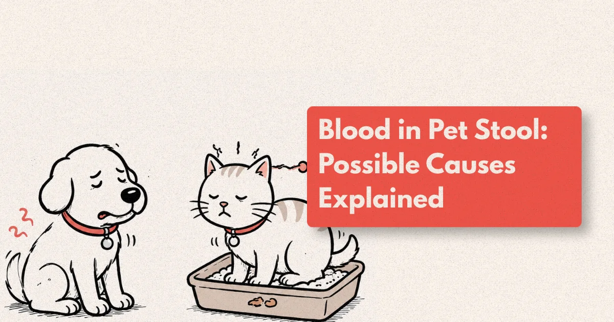
Finding blood in your dog’s or cat’s stool is alarming, and the alarm is sometimes warranted. Blood in pet stool ranges from a single episode caused by minor dietary indiscretion to a sign of intestinal obstruction, severe colitis, parvoviral enteritis, or a bleeding tumour. The challenge for owners is that there’s no visual appearance of blood that reliably separates the urgent from the non-urgent, and delay in recognising serious GI bleeding is one of the most common reasons a treatable condition becomes a complicated one.
Hematochezia (fresh red blood in or on stool) and melena (dark, tarry, digested blood originating from the upper GI tract) are among the most frequent presenting complaints in general veterinary practice, accounting for an estimated 12 to 15% of GI consultations annually (Journal of Veterinary Internal Medicine, 2025). The two have different anatomical origins, different differential diagnoses, and different levels of urgency. Understanding that distinction is the starting point for understanding what your vet is looking for when they begin a GI workup.
What to Know
Fresh red blood on or in stool (hematochezia) most commonly originates from the colon or rectum. Dark, tarry, foul-smelling stool (melena) indicates digested blood from the stomach or small intestine and is generally more serious. A single episode of hematochezia in a dog that is otherwise well, eating, and not lethargic warrants a veterinary call but not always an emergency visit; melena, bloody diarrhoea combined with lethargy or vomiting, or blood in stool in a puppy or unvaccinated dog should be assessed the same day.
What Does Blood in Pet Stool Actually Look Like?
The appearance and character of blood in stool provides the first and most important piece of diagnostic information before any test is run, because it tells you where in the gastrointestinal tract the bleeding is originating.
Hematochezia is fresh, bright red blood. It appears on the surface of formed or loose stool, mixed through soft stool, or passed as blood with mucus and little faecal matter. The bright red colour indicates that the blood has not had time to be digested, meaning the bleeding source is in the large intestine (colon, cecum) or the rectum and anus. Causes of hematochezia range from benign and self-limiting (mild colitis from dietary indiscretion, stress colitis, anal gland disease) to serious (colonic intussusception, polyposis, invasive colonic neoplasia, haemorrhagic gastroenteritis).
Melena is dark, tarry, sticky, foul-smelling stool that looks almost black and has a characteristic tar-like consistency. The dark colour comes from haemoglobin that has been digested as it transits through the small intestine and colon. It indicates bleeding originating from the oesophagus, stomach, or duodenum. The volume of blood required to produce visible melena is substantial, often 50 to 100 mL or more in a medium-sized dog, which means melena usually indicates significant and ongoing upper GI haemorrhage. Causes include gastric and duodenal ulcers (often secondary to NSAIDs, corticosteroids, or stress), gastric neoplasia, a swallowed foreign body eroding mucosa, and severe inflammatory bowel disease.
The distinction is clinical but imperfect: in dogs with very rapid lower GI transit, blood from a colonic source can pass quickly enough to remain bright red. Conversely, very slow upper GI transit can cause some darkening of blood from a more distal source. When the character of the blood is ambiguous, the rest of the clinical picture, including signalment, history, and associated signs, guides the interpretation.
What Causes Blood in Dog or Cat Stool?
The causes of GI bleeding in pets are divided by location (upper vs. lower GI) and by mechanism (mucosal inflammation, ulceration, vascular disruption, neoplasia, coagulopathy). Working through the differential systematically rather than treating empirically produces better outcomes, fewer relapses, and avoids the masking of serious underlying disease with symptomatic medication.
Lower GI Bleeding (Hematochezia)
Dietary indiscretion and acute colitis is the most common cause of a single, isolated episode of hematochezia in otherwise healthy adult dogs. A dog that has eaten an unusual food, raided the rubbish bin, or been under significant environmental stress (boarding, travel, a new pet or person in the household) will sometimes develop transient colitis producing loose stool with fresh blood and mucus. The blood is typically a small amount coating otherwise normal stool, the dog continues to eat and drink, and the episode resolves within 24 to 48 hours. This pattern accounts for approximately 35 to 40% of hematochezia presentations in general practice (JVIM, 2025).
Haemorrhagic gastroenteritis (HGE), now often termed acute haemorrhagic diarrhoea syndrome (AHDS), presents dramatically: sudden-onset profuse, bloody diarrhoea, often described as “raspberry jam” in appearance, with a packed cell volume (PCV) that rises rapidly due to fluid loss from the gut. It predominantly affects small-breed dogs and resolves quickly with aggressive IV fluid therapy, but can be life-threatening through hypovolaemic shock if untreated. The cause remains incompletely understood but appears linked to Clostridium perfringens toxin production and altered mucosal permeability.
Parasites. Hookworms (Ancylostoma and Uncinaria species) attach to the intestinal mucosa and feed on blood, producing bloody diarrhoea particularly in puppies and immunocompromised dogs. Whipworms (Trichuris vulpis) infest the cecum and large intestine and are a common cause of chronic hematochezia in dogs with outdoor exposure. Giardia and Tritrichomonas produce watery, mucoid diarrhoea and sometimes hematochezia in cats.
Parvovirus. In unvaccinated or incompletely vaccinated puppies and young dogs, canine parvovirus (CPV-2) produces severe haemorrhagic enteritis that is rapidly life-threatening. The stool is profusely haemorrhagic, often with a characteristic sweet, foul odour, and accompanied by profound lethargy, vomiting, and fever. Mortality without aggressive supportive treatment reaches 50 to 80% in severe cases. Parvovirus is the reason blood in stool in an unvaccinated or young dog is always urgent.
Colorectal polyps and neoplasia. Benign rectal polyps in dogs, particularly middle-aged to older Miniature Schnauzers and other small breeds, produce chronic intermittent hematochezia with formed stool, often with straining. Colorectal carcinoma and lymphoma produce similar signs but with progressive weight loss, tenesmus, and change in stool character over time. Digital rectal examination detects many rectal masses directly. A mass felt on rectal exam in a dog with hematochezia requires urgent workup.
Anal sac disease. Impacted or infected anal glands can produce blood-tinged material expressed during defecation and visible on or near the stool. This is often misidentified as hematochezia; examination of the perineum and anal glands during the physical exam distinguishes it.
Upper GI Bleeding (Melena)
NSAID and corticosteroid-induced gastropathy is the most common cause of gastrointestinal ulceration in dogs. Both drug classes impair the prostaglandin-mediated protective mechanisms of the gastric mucosa. Dogs receiving NSAIDs for orthopaedic pain management, or those on corticosteroids for inflammatory or immune-mediated conditions, carry elevated risk of gastric and duodenal ulceration, particularly when drugs from both classes are combined or when an NSAID is started after recent corticosteroid use. Melena in a dog on these medications is a gastrointestinal emergency until proven otherwise.
Gastric and intestinal foreign bodies. Swallowed sharp objects (bones, needles, wire, toys with hard edges) can erode through mucosal and submucosal layers during transit, producing haemorrhage at the point of lodgement or perforation. Haemorrhagic signs combined with anorexia and abdominal pain are red flags for a perforating or obstructing foreign body.
Neoplasia. Gastric adenocarcinoma, intestinal lymphoma, leiomyoma, and leiomyosarcoma all produce chronic upper or lower GI bleeding. The clinical pattern is often insidious: gradual weight loss, intermittent vomiting or diarrhoea, and progressive melena or hematochezia over weeks before the primary lesion is identified on imaging.
Systemic coagulopathies. Rodenticide toxicity (anticoagulant bait ingestion), immune-mediated thrombocytopenia, disseminated intravascular coagulation (DIC), and von Willebrand disease can all produce GI bleeding as part of a broader haemorrhagic syndrome. The clue is haemorrhage at multiple sites: petechiae on gums, subcutaneous bruising, haematuria, alongside GI blood loss.
Inflammatory bowel disease (IBD) and protein-losing enteropathy (PLE). Chronic IBD in both dogs and cats produces a variable combination of vomiting, diarrhoea, weight loss, and intermittent blood in stool. PLE occurs when severe intestinal inflammation disrupts the mucosal barrier to the point of protein leakage into the intestinal lumen; hypoalbuminaemia, oedema, and ascites can result in severe cases.
The practical takeaway from this distribution is that in adult, vaccinated dogs, the most common cause of a single episode of blood in stool is benign and self-resolving. But the tail of the distribution matters: every presentation that persists, recurs, or is accompanied by systemic signs is carrying the prior probability of a serious cause that requires investigation, not repeated empirical treatment.
Which Signs Mean Blood in Stool Needs Immediate Attention?
The character and volume of the blood, and what’s happening to the pet alongside it, determines the urgency tier.
Go to an emergency clinic the same day or immediately if:
- The blood in stool is accompanied by vomiting, severe lethargy, collapse, or pale gums. This combination suggests significant systemic illness or haemodynamic compromise.
- The stool is profusely haemorrhagic, like raspberry jam consistency throughout. This pattern characterises AHDS/HGE, which can cause hypovolaemic shock within hours.
- The pet is a puppy, is unvaccinated, or has unknown vaccination status. Parvoviral enteritis progresses rapidly and is lethal without IV fluids and supportive care.
- The stool is dark, tarry, and foul-smelling (melena) and the dog is on NSAIDs or corticosteroids. NSAID-induced ulceration can perforate, and perforation carries very high mortality.
- Multiple episodes of haemorrhagic stool in 24 hours with visible deterioration in the pet’s condition.
- Stool contains blood and the pet is known to have ingested rodenticide bait, grapes, raisins, or a sharp foreign object.
- Bruising on the skin, bleeding gums, or blood in the urine alongside bloody stool. This pattern suggests a systemic coagulopathy.
Call your vet for a same-day appointment if:
- A single episode of hematochezia in an adult, vaccinated, otherwise well dog that is still eating, active, and not lethargic. This pattern is most often stress or dietary colitis and can often be assessed non-urgently if the dog’s general condition is normal, but it still needs a vet call to confirm.
- Recurrent episodes of blood in stool over several days, even if the dog seems well between episodes.
- Blood in stool in a cat, regardless of volume. Cats rarely show GI signs unless the disease is significant.
- Blood in stool alongside progressive weight loss, vomiting, or appetite changes over days to weeks.
Melena always warrants same-day professional evaluation because the conditions it represents (upper GI ulceration, neoplasia, coagulopathy) are almost never benign and often require rapid intervention to prevent progression. A single episode of small-volume fresh hematochezia with normal systemic signs is the one pattern where a 24-hour monitoring window before calling may be reasonable in an adult dog with no complicating history.
How Is the Cause of GI Bleeding Diagnosed?
The diagnostic approach to blood in pet stool follows the same principle as respiratory triage: identify the location first, then characterise the cause. The history and physical examination give the location (upper vs. lower GI); laboratory and imaging workup defines the cause.
History and physical examination. The vet will ask about the character and duration of the blood, dietary history (recent food changes, scavenging, known toxin exposure), medication history (NSAIDs, steroids, any recent prescription changes), vaccination status, travel history, and other household pets. Physical examination includes abdominal palpation (pain, masses, gas distension), rectal examination (rectal masses, anal gland disease, stool character at source), and assessment of systemic signs (mucous membrane colour, hydration, heart rate, temperature).
Faecal examination. Direct faecal smear, faecal flotation, and faecal PCR panels are the first-line laboratory tests for GI bleeding cases. They detect endoparasites (hookworm, whipworm, roundworm eggs, Giardia cysts), and PCR panels expand this to include parvovirus, Clostridium, Campylobacter, Salmonella, and Cryptosporidium in a single submission. Parvovirus point-of-care ELISA testing on faeces gives results in 10 minutes and is performed immediately on any unvaccinated or young dog with haemorrhagic diarrhoea.
Complete blood count (CBC) and biochemistry panel. The CBC identifies anaemia (which quantifies the blood loss), thrombocytopenia (platelet deficiency causing failure of primary haemostasis), and inflammatory leukograms suggesting infection or significant tissue inflammation. The biochemistry panel assesses protein levels (hypoalbuminaemia suggests PLE or severe hepatic disease), renal and hepatic function, and electrolyte status. A markedly elevated PCV (>55–60%) with normal total protein is the signature of AHDS, distinguishing it from septic or haemorrhagic shock from other causes. Coagulation tests (PT, aPTT) are added when anticoagulant rodenticide toxicity or DIC is suspected.
Abdominal ultrasound. Ultrasound is the most valuable single imaging tool for GI bleeding investigation because it gives real-time information about all layers of the GI wall and the surrounding structures that radiographs cannot provide. A trained ultrasonographer can assess bowel wall thickness and layering (preserved layering suggests IBD or enteritis; lost layering suggests neoplasia), identify focal masses or thickening, detect intussusception, free abdominal fluid, and enlarged lymph nodes, and characterise foreign bodies. Ultrasound-guided fine needle aspirates (FNA) of mesenteric lymph nodes or focal masses can be obtained at the same examination, providing cytological information without the morbidity of surgery or anaesthesia.
Normal small intestinal wall thickness in dogs is 2 to 5 mm depending on body size; normal large intestinal wall thickness is 2 to 4 mm. Diffuse wall thickening with preserved layering across multiple bowel segments is highly consistent with IBD or lymphocytic-plasmacytic enteritis. Focal, asymmetric thickening with disrupted wall layering has a positive predictive value of approximately 65 to 75% for neoplasia in dogs and warrants biopsy for definitive diagnosis (Veterinary Radiology and Ultrasound, 2025).
Thoracic and abdominal radiographs. Radiographs remain important for identifying GI foreign bodies (particularly radio-opaque objects), assessing for signs of perforation (free abdominal gas on plain radiographs is a peritonitis emergency), evaluating gastric distension patterns, and screening for pulmonary or hepatic changes that may suggest metastatic disease. Where obstruction is suspected, contrast studies can outline the level and degree of the blockage.
Endoscopy and biopsy. When imaging suggests mucosal disease (IBD, neoplasia) without identifying a focal lesion amenable to surgical removal, endoscopy provides direct visualisation of the mucosa and allows targeted biopsy. Gastroduodenoscopy accesses the stomach and duodenum; colonoscopy accesses the colon and ileum. Tissue biopsy with histopathology is the only definitive way to distinguish IBD from intestinal lymphoma, a distinction that is clinically critical because the treatment is entirely different. In some cases, full-thickness intestinal biopsy via laparoscopy or surgery is required when endoscopic biopsies are superficial and non-diagnostic.
Abdominal ultrasound represents the inflection point in GI diagnostics: it moves the working diagnosis rate from 58% (achievable with history, physical examination, and laboratory testing) to 77%, and it guides whether the next step is medical management, endoscopy, or surgery. Delaying ultrasound in favour of additional rounds of empirical medication delays reaching that inflection point and extends the period during which the underlying disease continues.
How Is Blood in Pet Stool Treated?
Treatment is directed at the cause and cannot be meaningfully described without a diagnosis. This is one of the most important messages in GI medicine: empirical treatment of bloody stool without a diagnosis is appropriate only as a short-term measure while a diagnosis is being established, not as a substitute for establishing one.
Fluid therapy. Dogs with AHDS/HGE, profuse bloody diarrhoea, or any degree of clinical dehydration require IV fluid replacement. The haemoconcentration characteristic of AHDS means plasma volume is reduced even as PCV appears normal or elevated; aggressive fluid replacement is the cornerstone of treatment and the reason survival rates for AHDS managed promptly approach 95% (JVIM, 2025).
Dietary management. A short-term (48 to 72-hour) switch to a highly digestible, low-residue, or intestinal-formula diet reduces colonic fermentation load and mucosal irritation during acute colitis episodes. Dogs with confirmed food-responsive enteropathy require longer-term dietary management with novel protein or hydrolysed protein diets. For some dogs, diet alone (without additional medication) is sufficient to achieve full clinical remission from IBD; a subset of cases diagnosed as steroid-responsive IBD in the past are now recognised as food-responsive.
Antiparasitic treatment. Confirmed or strongly suspected endoparasite burdens are treated specifically: fenbendazole for hookworm, whipworm, and roundworm; praziquantel for tapeworms; metronidazole or fenbendazole for Giardia; ronidazole for Tritrichomonas in cats. Empirical broad-spectrum deworming is reasonable while awaiting faecal results in puppies and dogs with outdoor exposure.
Acid suppression and mucosal protection for upper GI bleeding. Dogs with gastric or duodenal ulceration receive proton pump inhibitors (omeprazole, pantoprazole) to reduce gastric acid production and allow mucosal healing. Sucralfate is used as an adjunct mucosal protectant, particularly for erosive oesophageal or gastric disease. Dogs on NSAIDs that develop GI bleeding have their NSAID discontinued (or switched to a more GI-selective option) and receive acid suppression therapy.
Immunosuppressive therapy for IBD. Confirmed inflammatory bowel disease that does not respond to dietary management is treated with corticosteroids (prednisolone, initially at immunosuppressive doses) with or without additional steroid-sparing agents (chlorambucil, cyclosporine) depending on severity and disease type. Low-grade alimentary lymphoma, which histologically can be difficult to distinguish from IBD, is treated with prednisolone and chlorambucil, a combination that achieves median survival times of approximately 23 months in cats and 18 months in dogs from the time of diagnosis (JVIM, 2025).
Surgical and endoscopic intervention. Intussusception requires surgical reduction or resection. Obstructing foreign bodies require surgical retrieval when endoscopic extraction is not possible or not safe. Resectable focal GI tumours (leiomyoma, low-grade focal carcinoma) are treated surgically. The decision between surgery, endoscopy, and medical management for GI neoplasia is made on the basis of disease type, extent, location, and surgical risk assessment.
What to Expect from a GI Diagnostics Appointment
A first GI consultation for a dog or cat with blood in stool typically takes 30 to 60 minutes including examination and in-house diagnostics. If abdominal ultrasound is indicated, it is often performed at the same visit. Faecal and laboratory results from external panels typically return within 24 to 48 hours.
Our internal medicine and GI diagnostics service includes in-house abdominal ultrasound with same-day reporting, point-of-care laboratory testing, faecal PCR submission, and direct referral pathways to endoscopy and surgical services for cases requiring further investigation. Where possible, we aim to have a working diagnosis and initial treatment plan at the end of the first consultation.
Frequently Asked Questions
Is a small amount of blood in my dog's stool always serious?
A single, small episode of fresh blood on otherwise normal stool in an adult, vaccinated dog that is eating, drinking, and not lethargic allows a 24-hour monitoring window before calling the vet. If the episode repeats, blood volume increases, or the dog’s condition changes, veterinary assessment is needed. In cats, puppies, unvaccinated dogs, and dogs on NSAIDs or steroids, even a single episode warrants a same-day call.
What does dark, black, tarry stool mean in dogs?
Dark, tarry, foul-smelling stool (melena) indicates digested blood from the upper GI tract (oesophagus, stomach, small intestine), signalling more significant haemorrhage than fresh hematochezia. It warrants same-day veterinary assessment. Common causes include NSAID-induced gastric ulceration, GI neoplasia, foreign body erosion, and coagulopathies. Certain medications (iron supplements, activated charcoal) and foods can darken stool without blood; true melena has a characteristic tar-like consistency and foul odour.
Can worms cause blood in my dog's stool?
Yes. Hookworms feed on intestinal mucosa and cause significant hematochezia, particularly in puppies. Whipworms infest the cecum and large intestine and are a common cause of chronic hematochezia in adult dogs. Annual faecal testing is recommended even in dogs on monthly preventatives, because no single product covers all intestinal parasite species (ESCCAP, 2025).
How is abdominal ultrasound used to investigate GI bleeding?
Abdominal ultrasound allows real-time assessment of bowel wall thickness and layering across all GI sections. It detects focal masses, diffuse thickening, intussusception, abnormal lymph nodes, free fluid, and foreign bodies. In dogs with GI bleeding, ultrasound moves the working diagnosis rate from approximately 58% (history and labs alone) to 77%, making it the highest-yield single imaging step. Ultrasound-guided fine needle aspirates can add cytological data at the same appointment (VRU, 2025).
My dog has had blood in the stool twice in a month but seems fine. Is that concerning?
Yes. Recurrent hematochezia, even in a dog that seems well between episodes, warrants investigation. Common underlying causes include colonic IBD, polyposis, low-grade parasitic burden, or food-responsive enteropathy — all of which have effective treatments once identified. Repeated empirical metronidazole courses without investigation delays diagnosis and allows disease progression. Faecal exam, bloodwork, and abdominal ultrasound at the second episode is the appropriate approach.