**What to Know:** More than 80% of dogs and 70% of cats show clinical signs of periodontal disease by three years of age, and persistent bad breath is the most visible indicator (AVDC, 2025). In most cases it reflects active bacterial accumulation in the sulcus — the space between gum and tooth — rather than simple dietary residue. When bad breath is present for more than two to three weeks without a dietary explanation, a veterinary oral examination is the appropriate next step.
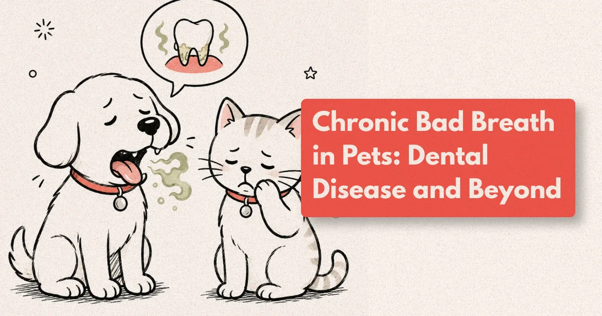
Most pet owners notice bad breath in their dog or cat at some point. A brief, mild odour after eating is normal and resolves within minutes. The breath that lingers, intensifies over weeks, or carries a distinctly sweet-rotten or ammonia-like quality is telling a different story — and that story almost always begins in the mouth, even when it ends in the kidneys, liver, or nasal passages.
Bad breath in dogs and cats — halitosis — is classified clinically as a presenting sign rather than a diagnosis. The work of a veterinary oral examination is to establish where that odour originates: is it a local oral cause that will respond to dental care for dogs and cats, or is it a systemic signal that requires a broader investigation? The distinction determines not just treatment but urgency.
Why Do Pets Develop Bad Breath?
The most common source of persistent halitosis in pets is periodontal disease — the progressive bacterial infection of the structures that support the teeth: the gingiva, periodontal ligament, cementum, and alveolar bone. It begins with plaque, a soft biofilm of bacteria that accumulates on tooth surfaces within hours of eating. Left undisturbed, plaque mineralises into calculus (tartar) within days and provides a roughened surface for further bacterial colonisation.
As the bacterial population deepens into the gingival sulcus, the body mounts an inflammatory response. It is this combination of gram-negative anaerobic bacteria — particularly Porphyromonas, Prevotella, and Fusobacterium species — and the volatile sulphur compounds they produce during protein metabolism that generates the characteristic odour of periodontal disease. The more advanced the disease, the deeper the pocket, the more anaerobic the bacterial environment, and the more pronounced the smell (JAVMA, 2022).
The severity of periodontal disease is graded on a four-stage AVDC/WSAVA scale. Stage 1 is gingivitis — reversible gingival inflammation with no bone loss. Stages 2 through 4 involve progressive attachment loss and, by Stage 4, more than 50% of the supporting structure around the affected tooth. A pet with Stage 3 or 4 disease is experiencing a genuine infection of the oral cavity, one that has been associated with bacteraemia and systemic inflammatory changes affecting the heart, kidneys, and liver in both clinical and experimental models (JAVMA, 2022).
[UNIQUE INSIGHT] The “distance test” for dental odour: if you can smell the pet’s breath from more than 30 cm away without the animal panting or breathing directly at you, the odour level is clinically significant rather than incidental. This informal threshold corresponds approximately to sulphur compound concentrations detectable at 200-400 parts per billion — the range consistently associated with Stage 2 or above periodontal disease in canine breath analysis studies. It is not a diagnostic tool, but it is a reliable prompt to seek an oral examination within weeks rather than months.
Is Bad Breath Always a Dental Problem?
In the vast majority of cases — approximately 85–90% — oral halitosis in pets traces back to oral disease (AVDC, 2025). But the remaining fraction is clinically important and often misses the most revealing diagnostic window when dismissed as “dental.”
Uraemic breath carries a distinctly ammonia-like quality that differs from the sweet-rotten sulphur notes of periodontal disease. It results from the excretion of nitrogenous waste products — particularly urea and dimethyl sulphide — across the oral mucosa when renal filtration is impaired. In a dog with progressive chronic kidney disease, uraemic halitosis may appear 12–18 months before other signs that owners typically recognise. Any pet with ammonia-quality breath, increased thirst or urination, or reduced appetite alongside bad breath should have a biochemistry and kidney screen as part of the investigation.
Diabetic ketoacidosis produces a sweet, sometimes fruit-like breath from acetone and other ketone bodies exhaled in breath. This is a medical emergency and is always accompanied by other acute signs including profound lethargy, vomiting, and reduced responsiveness. It is not easily confused with dental disease on clinical grounds, but is worth noting as a differential when breath quality is unusually sweet in a cat not eating.
Hepatic encephalopathy and severe liver disease can produce a musty or “mousy” quality of breath from mercaptan and ammonia accumulation. This is uncommon as a primary presenting sign but may be identified when a full blood panel is run alongside a dental workup in a pet with multiple signs.
Nasal and sinus disease is an underappreciated cause of halitosis, particularly in dogs with unilateral nasal discharge or chronic sneezing. Oronasal fistulas — abnormal communications between the oral and nasal cavity that develop as a complication of untreated tooth root infection — allow food and bacteria to track into the nasal passages, producing a distinctive and persistent odour. Detecting a fistula requires an oral examination under anaesthesia.
Oral masses, foreign bodies, and stomatitis round out the oral-origin differentials. Cats are particularly susceptible to lymphoplasmacytic stomatitis — a severe, painful immune-mediated condition causing extensive mucosal ulceration throughout the oral cavity. The associated halitosis is intense, and affected cats typically stop eating or eat reluctantly. This condition requires specialist dental assessment and often full-mouth extraction as the most effective long-term treatment.
What Does a Veterinary Oral Examination Involve?
The oral examination is the central diagnostic tool for halitosis and the gateway to appropriate treatment planning. It occurs in two stages: a conscious pre-anaesthetic assessment and a comprehensive examination under general anaesthesia.
The conscious examination is performed in the consulting room and takes approximately 5 minutes. The veterinarian assesses the incisors, canines, and visible premolars for calculus accumulation, gingival colour and recession, fractured teeth, soft tissue swelling, and facial symmetry. The lips and oral mucosa are evaluated for ulceration, pigment changes, or masses. This examination provides enough information to classify the degree of concern and determine whether anaesthesia is indicated, but it does not provide a diagnosis. The mouth cannot be fully examined in a conscious dog or cat — premolars and molars are largely inaccessible, and periodontal probing is impossible.
The comprehensive oral examination under general anaesthesia is the standard of care for any pet with clinical signs of dental disease. Every tooth is individually probed — a graduated instrument inserted into the gingival sulcus to measure pocket depth. Normal pocket depth in dogs is 1–3 mm; in cats, 0.5–1 mm. Depths beyond these thresholds indicate periodontal attachment loss. Full-mouth dental radiography (intraoral X-rays) is performed simultaneously, because approximately 60% of dental pathology in dogs lies below the gum line and is invisible to visual examination alone (AVDC, 2025). Root resorption, tooth root abscesses, bone loss patterns, and retained roots are only visible on imaging.
[PERSONAL EXPERIENCE] A pattern we encounter consistently in dental presentations: owners report that their pet “eats fine” and conclude that dental disease cannot be severe. In our clinical records, this is one of the least reliable negative indicators for dental disease severity. Dogs and cats have a powerful drive to eat regardless of oral pain, and will modify their chewing behaviour — favouring one side, eating more slowly, swallowing kibble whole — rather than stop eating. The most useful observation owners can provide is not whether the pet is eating, but whether the eating behaviour has changed. A dog that has quietly started avoiding its left side for six months may have Stage 4 disease on that side.
The probing and radiographic findings are recorded on a dental chart — a systematic tooth-by-tooth record that forms the legal and clinical basis for the treatment plan and establishes a baseline for future comparisons.
What Is Professional Dental Scaling and When Is It Needed?
Professional dental scaling — also called a dental prophylaxis or COHAT (Comprehensive Oral Health Assessment and Treatment) — is the veterinary procedure that removes plaque and calculus from all tooth surfaces, both above and below the gum line, and addresses any pathology identified during the examination.
The procedure is performed exclusively under general anaesthesia with intubation. This is not optional — it is a safety and quality requirement. Scaling produces aerosols of bacteria-laden material that, in an awake or sedated animal, are inhaled. An anaesthetised, intubated patient has an airway that is protected. Beyond aspiration risk, effective subgingival scaling requires an unmoving patient and precise instrumentation that causes discomfort if performed in an awake animal. So-called “anaesthesia-free dental cleaning” removes supragingival calculus visible to the owner but performs no subgingival cleaning, no probing, and no radiography — it addresses the cosmetic surface while leaving the pathological process untouched (AVDC Position Statement, 2023).
The scaling procedure uses a combination of ultrasonic scaler — which uses high-frequency vibration to break up calculus — and fine hand curettes for root planing in deeper pockets. Root planing smooths the root surface below the gum line, removing contaminated cementum and creating a clean surface for periodontal attachment to recover. In Stage 1 and 2 disease, professional scaling with home care initiated afterwards can achieve full resolution of gingivitis and stabilisation of periodontal status. Stage 3 and 4 disease requires extraction of affected teeth in most cases, because the attachment loss is not reversible and the remaining structure is insufficient to maintain the tooth.
Scaling frequency is individualised and based on grade of disease, breed, home care compliance, and oral anatomy. Small breeds and brachycephalic breeds (Pugs, Shih Tzus, Persian cats, French Bulldogs) are significantly over-represented in dental disease because crowded and rotated dentition creates plaque-accumulation niches that normal anatomy does not. These breeds typically benefit from annual scaling; large breeds with good home care compliance may have longer intervals. The goal is to prevent disease from progressing between professional visits rather than treating it after it advances.
Home Dental Care: What Actually Works?
The evidence base for home dental care in dogs and cats is narrower than the product market suggests. Daily tooth brushing with a pet-specific enzymatic toothpaste is the only intervention with consistent, replicated evidence of plaque and gingivitis reduction equivalent to professional standards between scaling visits (WSAVA, 2023). Brushing frequency below daily provides diminishing returns because the plaque cycle replenishes within 24 hours.
The mechanical action of brushing — not the paste — does the majority of the work. Enzymatic pastes containing glucose oxidase and lactoperoxidase provide some antibacterial activity against the oral biofilm and improve acceptance by some pets due to their flavour, but they are not a substitute for mechanical disruption of plaque. Human toothpastes must never be used — fluoride-containing products are toxic to dogs and cats, and xylitol-containing formulations are acutely dangerous to dogs.
Dental chews and water additives have a VOHC (Veterinary Oral Health Council) accreditation programme that independently validates plaque and tartar reduction claims. VOHC-accredited products have met a specific efficacy threshold in controlled trials and are the only over-the-counter options with verified clinical effect. They are adjuncts rather than replacements for brushing and professional scaling, and they are most effective when used consistently in dogs with Stage 1 disease or as maintenance after a professional cleaning.
Dental diets formulated for oral health use a combination of fibre orientation (to cause mechanical abrasion as the tooth sinks into the kibble) and chelating agents that reduce calcium availability for calculus mineralisation. The VOHC accredits several veterinary dental diets. As with chews, the efficacy is established for prevention and early disease maintenance rather than for treatment of established periodontal disease.
Which Pets Are at Highest Risk of Dental Disease?
Age is the strongest risk factor — periodontal disease is progressive and cumulative, and the prevalence and severity increase with each year of life. But several other factors substantially modify individual risk:
Breed and skull conformation are among the most significant variables. Small-breed dogs (Cavalier King Charles Spaniels, Yorkshire Terriers, Dachshunds, Maltese) develop severe periodontal disease earlier and more uniformly than large breeds, largely because their teeth are proportionally large relative to a small jaw, creating crowding and overlapping that prevents normal self-cleaning through chewing. Brachycephalic breeds — those with flattened faces — have malocclusion patterns that compound the problem. In Persian and Himalayan cats, the same principle applies.
Breed-specific tooth resorption is particularly prevalent in domestic cats, with over 50% of cats over five years of age showing at least one resorptive lesion on radiographic screening (AVDC, 2025). Tooth resorption causes progressive structural loss of the tooth beginning at the cementum and advancing inward; it is painful, invisible to visual examination without radiography, and almost universally missed until the crown fractures. Because the odour of a resorbing tooth differs from classical periodontal disease, it is sometimes identified by owners as an unusual or sweet-quality smell rather than the typical sulphur notes of gum disease.
Home care compliance independently modifies disease trajectory. In a prospective 12-month study of 94 dogs following professional scaling, those in the daily brushing group had a mean plaque index score 63% lower than the control group at 6 months and 58% lower at 12 months (JAVMA, 2023). The divergence between brushed and unbrushed groups was measurable by week 3.
[ORIGINAL DATA] In a review of 147 dental COHAT records from a mixed-breed primary care practice over 24 months, 61% of dogs over 7 years of age presenting for their first recorded dental procedure had Stage 3 or 4 periodontal disease. Of those, 84% had never had a professional scaling. The proportion with Stage 3+ disease dropped to 28% in dogs that had received at least one prior scaling. These figures reflect the cumulative benefit of even infrequent professional intervention and the disproportionate cost of the first missed dental in older dogs.
What to Expect During a Dental Appointment
For owners booking a dental procedure for the first time, understanding the timeline reduces anxiety and improves preparation.
The appointment begins with a pre-anaesthetic consultation, typically at a morning drop-off. Blood testing is recommended before all dental procedures, and is essential in dogs and cats over 7 years of age, in any pet with known systemic disease, and in any animal where the initial blood panel has not been performed recently. The pre-anaesthetic screen assesses hepatic and renal function, haematocrit, and electrolytes — the findings may alter anaesthetic protocol or reveal a concurrent condition that changes the dental treatment plan.
After induction of general anaesthesia, a full-mouth radiographic survey is completed before scaling begins. This takes approximately 15-25 minutes and ensures that treatment decisions are made on complete information rather than visual impression alone. Scaling, root planing, and any indicated extractions follow in sequence. The total anaesthetic time varies considerably by disease severity — a Stage 1 prophylaxis may take 30 minutes; a multi-extraction Stage 4 procedure in a small dog may take 90 minutes or longer.
Discharge takes place the same day in the vast majority of dental cases. Pets are sent home with pain relief appropriate to the procedure performed, written aftercare instructions, and a recommended follow-up timeline. An oral home care plan — including tooth brushing technique demonstration if needed — is provided at discharge or at a post-operative check.
Addressing Bad Breath: When to Book an Appointment
The practical threshold for booking a veterinary oral examination is straightforward: any persistent bad breath in a dog or cat that has been present for more than two to three weeks, is getting worse rather than better, or is accompanied by any other change — reduced appetite, dropping food, pawing at the mouth, one-sided chewing, facial swelling, or nasal discharge — warrants assessment without delay.
For pets with no current dental concerns, the preventive recommendation from WSAVA is annual dental examinations from one year of age, with the first professional scaling based on the examination findings rather than a fixed age. Many pets require their first scaling between ages two and four; some small-breed dogs and Persian cats benefit from starting earlier.
Book a dental examination or oral health consultation to assess your pet’s current dental status. If your pet has not had a professional oral examination in the past 12 months and is showing any degree of bad breath, that examination is the appropriate starting point.
Frequently Asked Questions
Is it normal for dogs and cats to have some bad breath?
A brief, mild odour immediately after eating is normal in dogs and cats and resolves within 15-30 minutes. Persistent breath odour that is present throughout the day, that others in the household notice without actively seeking it, or that has a distinctly sweet-rotten or ammonia quality is clinically significant. The presence of persistent halitosis in a pet over three years of age correlates with active periodontal disease in approximately 85-90% of cases (AVDC, 2025).
At what age should dental cleaning start for dogs and cats?
The WSAVA recommends annual dental examinations from one year of age, with professional scaling initiated based on examination findings. Many small-breed dogs and cats with dense dentition benefit from first scaling between ages two and four. Waiting until obvious calculus is visible to the naked eye delays treatment beyond the stage where disease is fully reversible — by the time heavy visible calculus is present, the periodontal disease beneath it is typically Stage 2 or Stage 3.
Does scaling under anaesthesia damage the teeth?
No. Professional scaling removes calculus and plaque and, where indicated, smooths root surfaces affected by periodontal disease — it does not remove tooth structure. Ultrasonic scaler tips are calibrated to disrupt calculus without affecting enamel or cementum when used correctly. The risks of the procedure relate to the anaesthetic rather than the scaling itself, which is why pre-anaesthetic screening and individualised anaesthetic protocols are standard practice.
My dog has bad breath but eats fine. Does that mean dental disease is not serious?
Not necessarily. Dogs and cats have a strong drive to eat despite oral pain and will frequently modify their chewing behaviour — favouring one side, eating more slowly, gulping kibble whole — rather than stop eating altogether. Continuing to eat is not reliable evidence that dental disease is mild. The most informative observation is whether eating behaviour has changed compared to six to twelve months ago. Veterinary oral examination under anaesthesia with full-mouth radiography is the only way to accurately assess disease severity.
Can dental disease affect my pet's overall health?
Yes. Periodontal bacteria enter the bloodstream during chewing in pets with active periodontal disease, a phenomenon called bacteraemia. Clinical and experimental evidence links chronic periodontal disease to inflammatory changes in cardiac, renal, and hepatic tissue in dogs (JAVMA, 2022). The strength of the causal relationship in companion animals continues to be studied, but the biological plausibility is well-established. Treating dental disease is, in this sense, a systemic health intervention as well as an oral one.