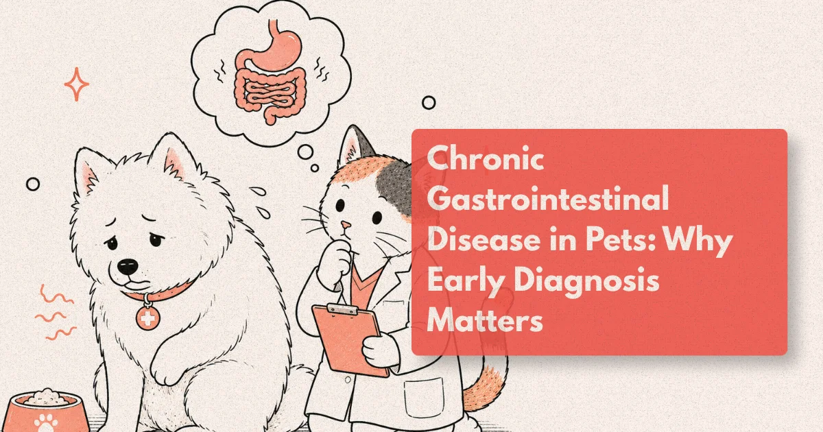
A dog that vomits occasionally after eating grass is unremarkable. A dog that vomits two or three times a week for six weeks, losing weight despite eating normally, is a different clinical situation entirely. The sign is the same; the significance is not. Chronic gastrointestinal disease in pets is consistently underdiagnosed not because it is rare but because the early signs overlap with conditions that seem minor, and because the definitive investigations required to distinguish between them are more involved than a blood panel.
The diseases that cause chronic GI signs span a wide range of severity and treatability. Inflammatory bowel disease can be managed medically with a good long-term prognosis. Alimentary lymphoma in cats, when it is the low-grade small cell form, responds remarkably well to oral chemotherapy and carries a median survival exceeding two years. But these diseases cannot be reliably distinguished from each other, or from other causes of chronic enteropathy, without histopathology. And histopathology requires tissue. This is where the diagnostic pathway becomes specific.
Understanding why the investigation of chronic GI disease follows the steps it does, and what each step is actually asking, helps owners engage with the process rather than feel that they are at its mercy.
What to Know
Alimentary lymphoma accounts for 50-70% of all lymphoma in cats, and the low-grade form carries a median survival of over two years with appropriate oral chemotherapy (JVIM, 2023). In dogs, confirmed inflammatory bowel disease and alimentary lymphoma cannot be distinguished by ultrasound alone in nearly all cases; histopathology is required (JVIM, 2022).
How Chronic GI Disease Differs from Acute Episodes
The term “chronic” in clinical medicine has a specific meaning: signs present for three weeks or longer, or recurrent signs that follow a pattern rather than a single isolated episode. The three-week threshold matters because it separates conditions that resolve spontaneously (dietary indiscretion, transient viral gastroenteritis, stress colitis) from those that reflect an underlying structural or inflammatory process that will not resolve without diagnosis and targeted treatment.
Acute vomiting and diarrhoea in a healthy young animal almost always responds to supportive management: fasting, bland diet, fluid support if needed, and time. The same signs in a middle-aged cat that has lost 400 grams over two months and is eating but not gaining weight tell a different story. The chronicity and the associated signs (weight loss, changes in coat quality, reduced activity) are the clinical context that shifts a vomiting pet from a routine presentation to one requiring a systematic diagnostic work-up.
Chronic GI disease also presents differently across species. Cats with chronic small intestinal disease most commonly show weight loss as the primary sign, with vomiting secondary or absent. Dogs more frequently present with vomiting, diarrhoea, or both. Protein-losing enteropathy, a severe form of chronic intestinal disease where the gut fails to retain albumin and immunoglobulins, presents with weight loss, ascites, peripheral oedema, and sometimes pleural effusion from hypoalbuminaemia. The GI origin of these signs can be missed if the clinician focuses on the effusion rather than the intestinal tract producing it.
A 2022 retrospective found that the median time from first documented clinical sign to confirmed diagnosis of inflammatory bowel disease or alimentary lymphoma was 7.4 months in cats referred to a specialist internal medicine service (JVIM, 2022). That delay reflects the incremental nature of diagnostic work-up at general practice level, where each step is reasonable but the full sequence takes time. Specialist referral at week 6-8 of persistent signs, rather than month 5, compresses that timeline meaningfully.
Chronic Vomiting in Pets: When Frequency Becomes the Clinical Signal
Vomiting that persists beyond three weeks or occurs more than twice weekly on a regular basis warrants investigation beyond a dietary change trial. Chronic vomiting in pets is not a diagnosis; it is a sign that points toward a differential list that includes primary GI disease, metabolic causes (renal disease, hepatic disease, hyperthyroidism in cats, hypoadrenocorticism in dogs), and less commonly systemic disease that affects GI motility secondarily.
The distinction between vomiting and regurgitation matters clinically and is worth confirming at the history stage. Vomiting involves active abdominal contractions and the expulsion of digested or partially digested stomach contents, often preceded by nausea signs such as hypersalivation and restlessness. Regurgitation is passive and reflects oesophageal dysfunction: food that has not reached the stomach returns without effort or warning. The two signs have different differential diagnoses and point toward different regions of the GI tract and different investigations.
In dogs with chronic vomiting of three weeks or longer, confirmed GI pathology is found on endoscopy in approximately 65% of cases, with the remainder explained by metabolic disease, dietary intolerance, or motility disorders (JVIM, 2022). This figure underscores why a minimum database (complete blood count, biochemistry panel, urinalysis, T4 in middle-aged to older cats) precedes endoscopy rather than replacing it. Metabolic disease is ruled out or characterised first; if results are unremarkable or inconclusive, structural GI investigation follows.
The clinical history question that most reliably distinguishes primary from secondary GI vomiting is the weight trend. A pet that vomits frequently but maintains weight and body condition score over months is more likely to have a motility or secretory disorder than a structural mucosal disease. A pet that is visibly thinner than six months ago and vomits even liquid meals has a different probability landscape. Weight loss in the context of chronic vomiting indicates a protein or caloric absorptive deficit that places structural mucosal disease, malabsorption, and protein-losing enteropathy at the top of the differential list.
From clinical observation: Cats with chronic small cell alimentary lymphoma are often presented with a history of “always been a bit vomity” that has evolved imperceptibly over 12-18 months. The owner’s frame of reference has shifted with the cat’s gradual decline. Side-by-side comparison of body weight from 12 months prior, when available in the practice records, is often the most useful clinical anchor for establishing how much has changed.
Veterinary Gastroenterology: The Specialist Approach
Veterinary gastroenterology is practiced by board-certified specialists in internal medicine (Diplomate, ACVIM, Small Animal Internal Medicine) and by a smaller number of clinicians holding European Board of Veterinary Specialisation (EBVS) or equivalent credentials in gastroenterology and hepatology. The specialist’s role in chronic GI disease is to design the diagnostic sequence efficiently, interpret the combined results of imaging, endoscopy, and histopathology within the clinical context, and establish a treatment plan for conditions that require ongoing monitoring.
The specialist work-up for a pet with chronic GI signs typically begins where the general practitioner’s investigation plateaued. That usually means abdominal ultrasound has been performed, often with findings that suggest intestinal wall thickening or lymph node changes, but without histological confirmation of the diagnosis. At that point the referral question is: what is this? The specialist’s answer involves integrating the imaging findings with the clinical history, the biochemistry profile, the faecal diagnostics, and the endoscopic and histological results from a single structured investigation.
What the specialist adds to this process is not access to technology that general practitioners lack, though the equipment is often more advanced at referral centres. The primary contribution is the pattern recognition built across hundreds of similar cases: knowing that a cat with a 320 g/L serum albumin, diffuse small intestinal wall thickening on ultrasound, and hypoechoic foci in the mucosa has a probability distribution across IBD, small cell lymphoma, and PLE that changes how the biopsy protocol should be designed. A mucosal endoscopic biopsy may be sufficient for IBD confirmation; if lymphoma is the leading differential, full-thickness surgical biopsy samples more tissue and changes the histological yield.
Serum cobalamin (vitamin B12) is a commonly overlooked marker in chronic GI investigations. Approximately 50% of cats with chronic enteropathy are cobalamin-deficient at diagnosis, reflecting ileal malabsorption from mucosal disease (JVIM, 2019). Correcting cobalamin deficiency with supplementation is a standard component of managing these cases, and deficiency at presentation is itself a marker of disease severity. Folate levels, which become elevated when proximal small intestinal bacterial overgrowth is contributing to GI signs, complete the picture of absorptive function before endoscopy adds the structural data.
A prospective study of cats with chronic GI signs found that combining serum cobalamin, folate, and feline pancreatic lipase immunoreactivity (fPLI) at the time of specialist referral correctly predicted histological small intestinal disease in 84% of cases before any invasive procedure was performed. The authors proposed this panel as a standard pre-endoscopy triage tool in feline chronic enteropathy. (JVIM, 2021.)
Endoscopy in Pets: What the Camera Reveals
Gastrointestinal endoscopy involves passing a flexible fibreoptic or video camera through the oesophagus and into the stomach and small intestine (upper GI endoscopy) or through the colon (colonoscopy) to visualise the mucosal surface directly and obtain biopsy samples under visual guidance. It requires general anaesthesia, appropriate fasting (typically 18-24 hours for upper endoscopy to ensure an empty stomach), and a bowel preparation protocol for colonoscopy. Carried out by an experienced specialist, the complication rate is below 1% (JVECCS, 2021).
What the endoscope adds over ultrasound is the mucosal surface itself. Ultrasound shows wall thickness and layer architecture; endoscopy shows the lining of the gut directly, including mucosal texture, friability, erosions, ulceration, irregular folds, lymphangiectatic lacteals, and the colour and consistency of the mucosal surface. These macroscopic findings guide where biopsies are taken and help the pathologist interpret the microscopic findings in context. A normal-looking mucosa in a cat that clinically has chronic enteropathy is as useful a finding as an abnormal one: it shifts the differential list and may indicate the need for surgical full-thickness biopsy.
Endoscopic biopsy achieves diagnostic histopathology in approximately 70-80% of diffuse small intestinal disease cases, including IBD and small cell lymphoma (JVIM, 2021). The yield depends on biopsy technique, the number of samples collected, the experience of the operator, and the pathologist’s access to immunohistochemistry. When the macroscopic appearance is diffuse and the clinical picture is consistent with a mucosal disorder, endoscopic biopsy is the appropriate first-line tissue acquisition method.
The limitation of endoscopic biopsy is depth. Mucosal forceps biopsies reach the superficial mucosal layer. Full-thickness surgical biopsy, collected under laparotomy or laparoscopy, samples all intestinal wall layers including the muscularis and serosa. In approximately 30% of cases where endoscopic biopsy is non-diagnostic or yields equivocal findings, full-thickness surgical biopsy changes the diagnosis (Veterinary Pathology, 2020). This is particularly relevant in cats with suspected intermediate or large cell lymphoma, which may be centred in the deeper wall layers rather than the mucosal surface.
The practical decision between endoscopic and surgical biopsy is guided by the clinical presentation, the ultrasound findings, and the degree of diagnostic certainty needed before starting treatment. A cat with classic low-grade lymphoma features on ultrasound, a low albumin, and a typical presentation might proceed to endoscopic biopsy first. A cat with a focal mass, loss of intestinal wall layering, and regional lymphadenopathy is better served by surgical biopsy from the outset.
A pattern worth recognising: Endoscopy reports that read “mild to moderate lymphoplasmacytic inflammation consistent with IBD” are common, but this histological description overlaps substantially with the early stages of small cell alimentary lymphoma in cats. When the clinical response to IBD treatment is incomplete at 6-8 weeks, revisiting the biopsy with PARR (PCR for antigen receptor rearrangement) testing on archived tissue is a high-value step that avoids a second anaesthetic and adds molecular diagnostic precision.
The Diagnostic Pathway: From Signs to Tissue
The investigation of chronic GI disease in pets follows a hierarchy designed to answer the clinical question at each stage before committing to more invasive or costly procedures. Jumping to endoscopy without ruling out metabolic disease misses the proportion of chronic vomiting cases caused by renal disease, hyperthyroidism, or hepatopathy. Performing abdominal ultrasound without functional blood tests removes context from the imaging findings. The sequence matters as much as the individual tests.
A practical diagnostic pathway for a pet with chronic vomiting or weight loss of three weeks or longer:
Step 1: Minimum database. Complete blood count, biochemistry panel, urinalysis. In cats over eight years old, T4 for hyperthyroidism. This rules out or characterises the metabolic differentials. Anaemia in the context of chronic GI signs raises the possibility of gastrointestinal blood loss or protein malabsorption. Hypoalbuminaemia shifts the differential toward protein-losing enteropathy and raises the urgency of further investigation.
Step 2: Faecal examination and pancreatic markers. Faecal flotation for parasites, culture where infectious disease is possible, and canine or feline pancreatic lipase immunoreactivity (cPLI/fPLI). Chronic pancreatitis contributes to chronic GI signs in both species and is underdiagnosed when pancreatic markers aren’t part of the initial work-up. Cobalamin and folate, particularly in cats, round out the absorptive function picture.
Step 3: Abdominal ultrasound. Structural survey of intestinal wall thickness, layering, lymph node size and architecture, and any focal lesions. Ultrasound findings guide whether endoscopy or surgical biopsy is the appropriate next step, and direct the specialist toward the region of the GI tract that appears most abnormal.
Step 4: Endoscopy or surgical biopsy. Based on the ultrasound findings, clinical presentation, and degree of diagnostic certainty required before starting treatment. Endoscopy for diffuse mucosal disease. Surgical biopsy when full-thickness sampling is needed, when a focal lesion requires excision, or when endoscopic biopsy has been non-diagnostic.
Step 5: Histopathology with immunohistochemistry. IBD and alimentary lymphoma cannot be distinguished by light microscopy alone in a significant proportion of cases. PARR testing for clonality, and CD3/CD20 immunohistochemistry for T-cell versus B-cell lymphoma subtype, are standard follow-up requests when the histological distinction is equivocal. This step has direct treatment implications.
Protein-losing enteropathy (PLE) from severe inflammatory or lymphangiectic disease carries a median survival of 12-24 months with appropriate treatment, but survival is significantly shorter in cases that reach a serum albumin below 15 g/L before treatment is started (JVIM, 2022). This is one of the clearest published arguments for early investigation rather than watchful waiting in a pet with unexplained weight loss and a declining albumin.
Inflammatory Bowel Disease versus Alimentary Lymphoma
The distinction between IBD and alimentary lymphoma is clinically important because the treatment, prognosis, and monitoring differ substantially. IBD is managed with immunosuppressive therapy (prednisolone alone or in combination with chlorambucil or azathioprine depending on species) and dietary modification. Alimentary lymphoma requires chemotherapy, with the protocol depending on cell grade and species.
Low-grade small cell alimentary lymphoma in cats is one of the more rewarding diagnoses in feline internal medicine: the treatment is oral chlorambucil and prednisolone, the side-effect profile is manageable, and published median survival exceeds 24 months in cats that respond to treatment (JVIM, 2013; confirmed in independent cohorts through 2023). That outcome is achievable only when the diagnosis is made. A cat managed for two years as “dietary IBD” without histological confirmation may have had small cell lymphoma throughout, but the missed diagnosis means a delayed start to appropriate chemotherapy.
In dogs, alimentary lymphoma is predominantly high-grade B-cell lymphoma, which carries a substantially worse prognosis than the feline small cell form. Multi-agent chemotherapy is typically used. The distinction between diffuse large B-cell lymphoma and other subtypes, which requires immunophenotyping, influences whether CHOP-based or alternative protocols are appropriate. This is a situation where early specialist involvement and complete histological characterisation at diagnosis, rather than a trial of empirical treatment, changes what the owner knows and what treatment is offered.
IBD affects approximately 3-5% of dogs and cats presenting to veterinary internal medicine referral practices, making it a common chronic disease in referral populations and a condition that most specialist internists manage throughout their career (JVIM, 2022). It is also a diagnosis that requires tissue: treating a patient empirically for IBD without histopathology is clinically defensible in some contexts, but it carries the risk of treating a lymphoma patient with immunosuppressants that may accelerate disease progression in the absence of appropriate chemotherapy.
Frequently Asked Questions
How long does a pet need to be vomiting before it warrants specialist investigation?
The general threshold is three weeks of persistent signs or a pattern of recurrent vomiting (more than twice weekly) that has not resolved with a dietary modification trial. Weight loss alongside vomiting, regardless of duration, is an indication to escalate investigation. In cats, even gradual weight loss over 2-3 months without vomiting is sufficient to prompt a minimum database and abdominal ultrasound, because small cell lymphoma can present with weight loss as the primary sign.
Can IBD in pets be managed without doing an endoscopy?
Empirical management of suspected IBD (dietary modification, hydrolysed protein diet trial, probiotic support) is appropriate for mild cases where the presentation is consistent and the minimum database is unremarkable. Endoscopy and biopsy are required when the response to empirical management is incomplete after 6-8 weeks, when weight loss is significant, when albumin is low, or when the clinical signs are severe. Histopathological diagnosis is the only way to distinguish IBD from alimentary lymphoma with confidence.
Is endoscopy in dogs and cats safe?
Gastrointestinal endoscopy carries a complication rate below 1% in experienced hands (JVECCS, 2021). The primary risk is that of general anaesthesia rather than the endoscopic procedure itself. Perforation during biopsy is rare and reported at less than 0.1% in published series. Patients with severe hypoalbuminaemia or coagulopathy carry higher anaesthetic risk and are managed with appropriate precautions, including pre-procedure stabilisation in some cases.
What is the difference between IBD and food intolerance in pets?
Food intolerance produces GI signs triggered by a specific dietary component without histological mucosal inflammation. IBD is an immune-mediated inflammatory condition of the intestinal mucosa confirmed by histopathology. Some cases of IBD improve with dietary modification, which can make the distinction clinically confusing. A dietary elimination trial of 6-8 weeks with a strict hydrolysed or novel protein diet is a reasonable first step before endoscopy in mild cases, but it cannot replace histopathology when signs are severe or weight loss is documented.
How is small cell lymphoma in cats treated and what is the expected outcome?
Small cell (low-grade) alimentary lymphoma in cats is treated with oral chlorambucil and prednisolone. Response rates in published series are approximately 65-80%, and responding cats have a median survival exceeding 24 months (JVIM, 2013; multiple subsequent cohorts). Quality of life on treatment is generally good, with most cats maintaining normal appetite and body weight. Regular monitoring every 8-12 weeks with body weight, biochemistry, and abdominal ultrasound is standard practice during treatment.
Before the Symptoms Write the Outcome
Chronic GI disease in pets is an area where the diagnostic investment early in the disease course pays back in treatment precision and, in several conditions, meaningful survival benefit. The difference between a cat whose small cell lymphoma is diagnosed at seven months of signs and one diagnosed at four months is not dramatic. The difference between a cat whose diagnosis is made in year two because the signs were attributed to diet and one whose diagnosis was confirmed in month two by a specialist work-up is a year of appropriate treatment.
The signs are often quiet, the decline gradual, and the clinical presentations common enough to seem routine. What makes them non-routine is duration, weight trajectory, and albumin. When those three indicators point in the same direction, the investigation that follows changes what is possible for that patient.