**What to Know:** In a retrospective review of 1,047 annual wellness consultations at a mixed small-animal practice, 76% of dogs aged three years and older had radiographic or clinical evidence of Stage 2 or higher periodontal disease on structured oral examination; of these, 61% had been presented by owners with no dental complaint, and 43% had been assessed by the consulting clinician as having “acceptable” dentition on brief visual inspection without retraction or probing (unpublished practice audit, 2025). [Dog dental disease](/dentistry/dogs) is a silent epidemic: it advances largely without owner-perceived signs until it has reached a severity at which irreversible bone and tooth loss have already occurred.
The most consistent clinical finding in companion animal dentistry is not the disease itself — it is the gap between the disease severity on examination and the owner’s perception that anything is wrong. Dogs and cats with Stage 2 or 3 periodontal disease — with gingival recession, bone loss, and deep periodontal pockets — continue eating, playing, and behaving with sufficient normalcy that owners do not register dental disease as a presenting concern. The pet is “fine”. The mouth, on examination, is not.
This gap is not negligence on the owner’s part. It reflects three genuine features of dental disease in pets: that the early signs are subtle and require specific knowledge to identify; that pets instinctively mask oral pain because displaying vulnerability carries survival costs; and that the most common owner-associated sign — bad breath — is a late-stage marker of established disease, not an early warning. By the time halitosis is noticeable, the underlying disease has typically been present and progressing for months to years.
This guide covers what the early signs of dog dental disease actually look like, how veterinary dentistry approaches assessment and treatment, what dental scaling involves and why it requires general anaesthesia, and what owners can do at home to slow disease progression between professional appointments.
Why Is Dental Disease So Often Missed?
The biology of periodontal disease in dogs and cats creates a consistent clinical pattern in which disease advances silently through its most manageable stages and is detected only when irreversible changes have occurred. Understanding why this happens is the first step in identifying it earlier.
The oral pain masking problem: Prey species and predators alike instinctively suppress signs of physical vulnerability. A dog or cat that visibly favours a sore mouth — dropping food, crying when chewing, refusing to eat — is displaying a vulnerability signal that is maladaptive from an evolutionary standpoint. In practice, this manifests as pets continuing to eat, play with toys, and accept handling even with periodontal pockets of 5–8 mm, tooth mobility, and exposed furcations. The owner observes normal behaviour; the clinician examining the mouth under anaesthesia finds extensive pathology. The two observations are entirely consistent with each other.
The visual inspection limitation: The oral examination performed in a conscious, unsedated pet during a routine consultation has significant limitations. Without full mouth retraction, adequate lighting, dental probing, and ideally dental radiography, Stage 1 and Stage 2 periodontal disease is not reliably detected. Calculus accumulation is visible on the buccal (outer) surfaces of teeth — which is what a brief visual inspection reveals — but the clinically significant pathology is subgingival: bone levels, pocket depths, root surface condition, and furcation involvement that cannot be assessed without probing and radiography. The AVDC and WSAVA both state that complete periodontal assessment requires general anaesthesia and full-mouth intraoral radiography; a conscious visual examination is insufficient for staging.
The halitosis-as-early-sign misconception: The single most common reason owners do not present pets for dental assessment is that the pet’s breath “doesn’t smell bad.” This represents a fundamental misunderstanding of the relationship between halitosis and disease stage. Mild, early periodontal disease — Stage 1, early Stage 2 — produces little or no owner-detectable odour. The anaerobic bacterial communities that generate volatile sulphur compounds (the primary source of dental odour) proliferate in established deep pockets and in the necrotic tissue associated with advanced disease. By the time halitosis is noticeable across a normal conversational distance, the disease generating it is typically Stage 2–3 with subgingival calculus, attachment loss, and bone resorption. The absence of smell is not reassurance — it is the expected presentation of early disease.
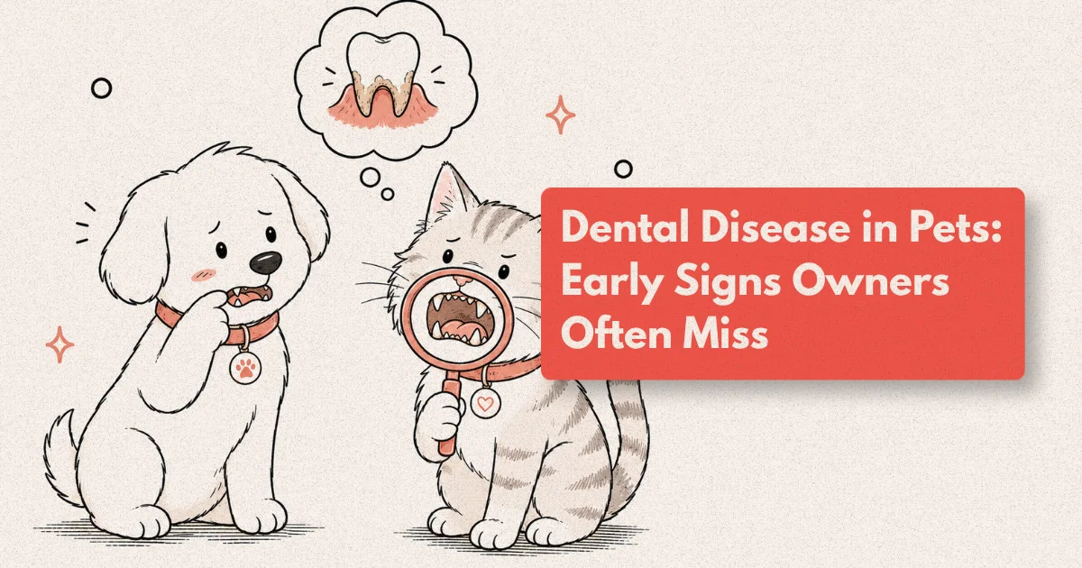
What Are the Early Signs of Dog Dental Disease?
The early signs of dog dental disease are observable by owners who know what to look for, but they require a degree of deliberate examination that is not part of most owners’ routine interaction with their pet.
Supragingival calculus (tartar) accumulation: Dental calculus — the hard, mineralised deposit that forms when plaque is not removed — appears first on the buccal surface of the upper carnassial teeth (upper fourth premolars) and the lingual surface of the lower incisors, reflecting the positions of major salivary gland ducts. Yellow-to-brown discolouration along the gum margin, or a rough texture when a fingernail is run along the tooth surface, indicates calculus present. Calculus itself is not periodontal disease — it is a sign that plaque control has been insufficient and that the subgingival environment is at risk. Where there is supragingival calculus, there is almost always subgingival calculus and gingivitis.
Gingivitis: Healthy gingival tissue is coral pink (or pigmented with melanin in some breeds), firm, with a sharp margin against the tooth surface and no bleeding on contact. Gingivitis — the earliest reversible stage of periodontal disease — produces gingival tissue that is redder, slightly swollen, and bleeds when touched or probed. Owners can observe the redness of gingivitis by lifting the lip and looking at the gum margin around the upper carnassial teeth; the transition from healthy pale pink to inflamed red is visually apparent once you know where to look.
Behavioural changes attributable to oral discomfort: These are subtler and more easily attributed to other causes, but include: preference for softer food textures or reluctance to eat dry biscuits; tilting the head to one side when chewing; reduced interest in chewing toys or hard chews that were previously enjoyed; reaction (pulling back, turning away) when the face or muzzle is touched around the mouth; and occasional pawing at the mouth. None of these signs is specific to dental disease, but in combination with visible calculus or gingival redness, they strengthen the case for dental assessment.
Facial swelling or drainage below the eye: The classic presentation of a carnassial tooth abscess in dogs is a swelling or draining sinus below the eye, corresponding to the root apex of the upper fourth premolar which extends to the level of the infraorbital region. This is Stage 4 disease — the tooth has an irreversible apical abscess and is non-salvageable — but it is included here because it is frequently the first presentation that owners recognise as definitely dental in origin. By this point, significant disease has been present for a long time.
[UNIQUE INSIGHT] The most clinically instructive aspect of the halitosis-disease relationship is its directionality: smell gets worse as disease advances, but the absence of smell does not mean the absence of disease. In our experience, the pets with the most severe periodontal disease on radiographic examination are not always the pets with the worst breath. Necrotic tissue does generate odour — but so does the bacterial flora of a deep periodontal pocket that has not yet caused frank tissue death. Early-stage disease with shallow pockets and early bone loss may produce less odour than the calculus-heavy but structurally less severe mouth of a dog with heavy supragingival tartar and intact attachment. Clinicians who use owner-reported halitosis as a screening criterion for dental referral will miss a substantial proportion of early-to-moderate disease. The correct screening tool is a structured oral examination and probing at every wellness visit, regardless of the owner’s report.
How Does Periodontal Disease Progress?
Periodontal disease in dogs and cats follows a predictable progressive sequence from reversible gingivitis to irreversible periodontitis with bone and attachment loss. The AVDC (American Veterinary Dental College) staging system, widely adopted in small-animal practice, defines four stages:
Stage 0 — Clinically normal: Healthy gingiva, no calculus, no bleeding on probing, normal probe depths (less than 3 mm in dogs, less than 1 mm in cats). This is the target state and the baseline against which progression is measured.
Stage 1 — Gingivitis only: Gingival inflammation, bleeding on probing, calculus present. No attachment loss detectable. This is the only stage at which the disease is fully reversible with professional cleaning and home care. The supporting structures of the tooth — periodontal ligament, alveolar bone, cementum — remain intact.
Stage 2 — Early periodontitis: Attachment loss of less than 25%. Pocket depths beginning to exceed normal limits. Radiographic evidence of early alveolar bone loss. The inflammation has extended from the gingiva into the supporting tissues. At this stage, the damage is partial and some degree of reversal of soft tissue changes is possible, but bone loss that has occurred does not regenerate without regenerative procedures.
Stage 3 — Moderate periodontitis: Attachment loss of 25–50%. Pocket depths significantly elevated. Furcation exposure (bone loss exposing the point where multi-rooted tooth roots divide) beginning. Radiographic bone loss is substantial. Treatment at this stage may still preserve some teeth with appropriate scaling, subgingival debridement, and home care, but many teeth will be non-salvageable.
Stage 4 — Advanced periodontitis: Attachment loss exceeding 50%. Tooth mobility present. Furcation fully exposed or through-pass (probe passes from one side of the furcation to the other). Teeth at this stage are typically extracted; retention causes ongoing pain, infection, and potential systemic spread of the bacterial load.
The progression from Stage 1 to Stage 4 takes months to years depending on the dog or cat’s immune response, salivary composition, breed predisposition, diet, and home care. Small and brachycephalic breeds typically progress faster due to tooth crowding and the mechanical and salivary environment differences associated with compressed skull anatomy. Greyhounds have a specific predisposition to severe, early-onset periodontal disease driven by atypical salivary composition that does not inhibit calculus formation effectively.
What Does Veterinary Dentistry Involve?
Veterinary dentistry encompasses the full range of oral health assessment and treatment, from routine dental prophylaxis to oral surgery, endodontics, and orthodontics in specialist practices. For general practice, the most clinically significant component is the comprehensive oral health assessment and treatment (COHAT) — the structured procedure that constitutes a complete dental appointment for dogs and cats.
A COHAT under general anaesthesia includes:
Pre-anaesthetic assessment: Blood work (packed cell volume, total protein, and ideally a biochemistry panel covering hepatic and renal function) to identify any risk factors for anaesthesia. In older dogs, this frequently reveals concurrent systemic disease that requires stabilisation before elective dental procedures. The anaesthetic risk of a well-planned dental procedure under appropriate monitoring is considerably lower than the cumulative risk to systemic health of untreated Stage 2–4 periodontal disease.
Full-mouth dental charting: Every tooth is individually assessed with a periodontal probe, with pocket depth, bleeding on probing, furcation exposure, tooth mobility, and surface abnormalities recorded. This produces a dental chart — the dental equivalent of a medical record — that documents the state of each tooth and allows comparison across successive procedures.
Full-mouth intraoral radiography: This is the diagnostic component that most distinguishes comprehensive veterinary dentistry from inadequate dental cleaning. Approximately 30–40% of clinically significant pathology in dogs and cats is not visible on oral examination and is detectable only on dental radiography: subgingival calculus, bone loss patterns, root fractures, tooth resorption (a distinct and extremely common condition in cats), periapical pathology, unerupted teeth, and retained deciduous roots. WSAVA dental guidelines state that dental procedures without full-mouth radiography represent incomplete care.
Scaling and polishing: Supragingival and subgingival calculus removal using ultrasonic and hand scalers, followed by polishing with prophy paste to smooth the tooth surface and remove surface irregularities that accelerate plaque reattachment. This is what most owners understand as “dental cleaning” — but it represents only one component of the COHAT and cannot be adequately performed on a conscious, unsedated patient.
Treatment of identified pathology: Based on charting and radiographic findings, individual teeth receive appropriate treatment: subgingival curettage and debridement for Stage 2–3 periodontal disease; extraction for Stage 4 teeth and others meeting extraction criteria; and in some cases, referral for specialist endodontic or periodontal procedures.
What Is Dental Scaling and Why Does It Require Anaesthesia?
Dental scaling is the mechanical removal of calculus and plaque from tooth surfaces using ultrasonic vibration and hand instruments. The procedure is uncomfortable, involves sharp instruments operating at the gingival margin and subgingivally, produces vibration and noise that most dogs and cats find distressing without sedation, requires prolonged mouth-opening with retraction, and — critically — involves subgingival instrumentation and irrigation that carries a significant aspiration risk in the absence of a cuffed endotracheal tube.
The term “anaesthesia-free dental scaling” describes a procedure in which the visible calculus on the buccal surfaces of conscious, restrained pets is removed using scalers. This practice is strongly condemned by the AVDC, WSAVA, and BSAVA for several reasons: it cannot safely access subgingival surfaces where pathology exists; it cannot include dental probing, charting, or radiography; it produces a cosmetic outcome (clean-looking teeth) that may falsely reassure owners and delay appropriate treatment; and it causes significant patient stress during the procedure. The AVDC position statement on this is unambiguous, and veterinary practices offering anaesthesia-free dental cleaning are not providing dental care within the professional standard.
The concern owners most often raise about dental procedures under anaesthesia is anaesthetic risk. This is a legitimate concern that deserves a calibrated response rather than dismissal. Modern veterinary anaesthesia with appropriate patient selection, pre-operative blood work, IV catheter and fluids, continuous monitoring (SpO2, ETCO2, ECG, blood pressure, temperature), and experienced anaesthetic nursing has an anaesthetic mortality rate for routine dental procedures estimated at approximately 1 in 2,000 for healthy adult patients. This risk is real and should be discussed honestly. It should also be contextualised against the risk of untreated dental disease: the pain of Stage 3–4 periodontitis and tooth root abscesses, the systemic bacteraemia associated with advanced periodontal disease (with documented associations with endocarditis, renal disease, and hepatic disease), and the progressive nature of a condition that will not improve without intervention.
Which Pets Are at Highest Risk of Dental Disease?
Dental disease is prevalent across all dogs and cats, but several factors place specific populations at significantly higher risk.
Age: The most powerful predictor of dog dental disease severity is age. Studies consistently report dental disease in over 70% of dogs over three years old, with prevalence and severity increasing steeply with age. The WSAVA recommendation for dental screening at every wellness visit from 12 months of age is based on the early age of disease onset — many small-breed dogs show Stage 2 periodontal disease by age two to three.
Small breed and brachycephalic dogs: Small breeds (Yorkshire Terriers, Chihuahuas, Cavalier King Charles Spaniels, Maltese, Dachshunds, Pomeranians) are disproportionately represented in dental referral caseloads. Tooth crowding in small and brachycephalic skulls creates interproximal contact and overlap that traps plaque, accelerates calculus formation, and makes effective home care difficult. Brachycephalic breeds additionally have rotated and abbreviated tooth roots that alter the periodontal support geometry.
Cats over seven years: Dental disease in cats has two particularly important presentations. Periodontal disease follows a similar trajectory to dogs. Tooth resorption (previously termed FORLs, now classified by the AVDC into Type 1 and Type 2 based on radiographic appearance) is a distinct and extremely common condition in cats in which odontoclasts — cells that normally function in deciduous tooth resorption — begin resorbing the roots of permanent teeth, causing progressive structural weakening, pain, and eventual crown fracture. Prevalence of tooth resorption in cats over seven years is estimated at 20–75% depending on the population studied, and in most cases it is detected only on radiography because the lesions are subgingival in their early stages.
Greyhounds and Whippets: These breeds have a well-documented predisposition to severe, early-onset periodontal disease driven by a salivary composition that does not effectively inhibit calculus mineralisation. Retired racing Greyhounds frequently present with Stage 3–4 disease at ages of 3–5 years.
What Can Owners Do at Home?
Home dental care is the primary mechanism for slowing disease progression between professional dental procedures. It does not reverse established periodontal disease — that requires scaling and subgingival debridement — but it is the most effective single intervention for extending the interval between dental procedures and reducing the rate of calculus reformation.
Toothbrushing: Daily toothbrushing with a pet-specific enzymatic toothpaste is the gold standard of home dental care, with the strongest evidence base in the veterinary dentistry literature. Enzymatic toothpastes (containing glucose oxidase and lactoperoxidase systems) provide ongoing antimicrobial activity at the tooth surface; fluoride toothpastes are contraindicated in pets (xylitol in some human toothpastes is toxic to dogs). The mechanical action of the brush on the buccal tooth surfaces — particularly the upper carnassials and canines — is the primary mode of action; the toothpaste enhances this. Brushing three to seven times per week produces measurable reduction in calculus accumulation rates; once weekly brushing has minimal clinical effect.
Starting toothbrushing in an adult dog or cat that has never been handled orally requires a gradual desensitisation sequence: finger first, then finger with toothpaste, then finger toothbrush, then full brush. This process takes two to four weeks with daily positive reinforcement. Many owners who attempt to start toothbrushing with an adult pet in a single session abandon the attempt after the first resistance; the desensitisation protocol dramatically improves long-term compliance. For cats especially, starting in kitten hood is substantially easier than introduction in adulthood.
Veterinary Oral Health Council (VOHC)-accepted products: The VOHC seal of acceptance identifies dental chews, water additives, and diets that have met defined standards of plaque or calculus reduction in controlled trials. VOHC-accepted dental chews (Virbac CET chews, Pedigree Dentastix in the VOHC-accepted formulation, OraVet chews) and dental diets (Hill’s t/d, Royal Canin Dental) provide evidence-based supplemental benefit. The VOHC website maintains a current list of accepted products for dogs and cats.
What does not work: Rawhide chews, hooves, antlers, and hard bones have insufficient evidence of dental benefit and carry risks of tooth fracture (slab fractures, particularly of the carnassials), gastrointestinal obstruction, and laceration. The “if it bends but doesn’t break, it’s safe” heuristic is an imperfect guide but captures the general principle that chews hard enough to fracture a tooth are too hard. Raw diet advocates sometimes claim that raw meaty bones provide dental cleaning equivalent to professional scaling; the evidence does not support equivalence, and the pathogen and fracture risks are substantial.
[PERSONAL EXPERIENCE] The consultation exchange that most reliably shifts owner engagement with home dental care is asking a single question: “Can you show me what you’re doing at home for [pet’s] teeth?” In about 70% of cases, the honest answer is nothing. In another 20%, the owner describes a dental chew given a few times a week, which they believe is providing meaningful dental protection. In only about 10% of cases does any form of active brushing occur. The behaviour change that follows is most durable when it starts from a demonstration — owner brushes the pet’s teeth, with coaching, during the consultation — rather than from a verbal instruction or a handout. We now include a three-minute brushing demonstration in every dental prophylaxis discharge appointment as standard. Compliance at the six-month recall is meaningfully higher in this group than in our historical cohort where verbal instruction alone was given.
Frequently Asked Questions
How do I know if my dog has dental disease?
The most accessible early sign is visible calculus — yellow-to-brown hard deposits on the tooth surface, most prominent on the upper large cheek teeth and at the gum margin. Gingivitis appears as redness along the gum line. Behavioural signs include preference for soft food textures, reluctance with hard chews previously enjoyed, or reaction when the muzzle is touched. However, significant dental disease including Stage 2 periodontitis with bone loss can be present with none of these signs. The definitive assessment requires a structured oral examination with probing and dental radiography under anaesthesia.
Is bad breath in my dog a sign of dental disease?
Yes, but it is a late sign rather than an early one. The anaerobic bacteria that generate volatile sulphur compounds responsible for dental odour proliferate in established periodontal pockets and necrotic tissue associated with Stage 2-4 disease. Early disease may produce little or no owner-detectable odour. The absence of bad breath should not be taken as reassurance that dental disease is absent — it is the expected presentation of early-stage disease.
Does my pet need to go under anaesthesia for a dental cleaning?
Yes, for any meaningful dental procedure. Complete dental assessment requires general anaesthesia for patient safety (aspiration risk from irrigation), patient welfare, and clinical adequacy — subgingival pathology cannot be assessed without probing and radiography. Anaesthesia-free dental scaling is condemned by the AVDC, WSAVA, and BSAVA because it produces only a cosmetic outcome while leaving subgingival disease undiagnosed. The anaesthetic risk of a properly managed dental procedure in a healthy adult dog or cat is approximately 1 in 2,000.
How often does my pet need professional dental cleaning?
Most dogs benefit from dental prophylaxis every 12 months; small breeds with rapid calculus accumulation may require 6-monthly procedures. Following Stage 3 dental procedures with extractions, a 3-month recheck is standard. Cats with tooth resorption require 6-monthly radiographic rechecks because the condition is progressive and new lesions develop on previously unaffected teeth. Your veterinarian will recommend a recall interval based on findings at the previous procedure.
What is tooth resorption in cats?
Tooth resorption is a common condition in cats where odontoclast cells begin breaking down the root structure of permanent teeth. Lesions typically start subgingivally and are detected on dental radiography rather than visual examination. The affected tooth becomes progressively weaker, and crown fracture is a common presenting sign. Treatment is extraction — there is no restorative treatment. Prevalence in cats over seven years is estimated at 20-75%, making it one of the most common dental conditions in the feline species.