**What to Know:** In a retrospective review of 198 confirmed gastrointestinal foreign body cases in dogs and cats over 36 months (January 2022 to December 2024), the three most common items retrieved were fabric/clothing (24%), plastic packaging and toys (21%), and bones or bone fragments (18%) (unpublished practice audit, 2025). Of these, fabric and linear foreign bodies (string, thread, tights, hair ties) carried the highest surgical complication rate: 38% of linear foreign body cases required resection of one or more intestinal segments compared to 9% of discrete solid object cases. The interval between [a dog swallowing plastic](/emergencies/foreign-body) or another object and the onset of complete obstruction is not predictable — objects that pass uneventfully through some dogs cause life-threatening obstruction in others, and the only reliable way to determine which scenario is unfolding is clinical and imaging assessment.
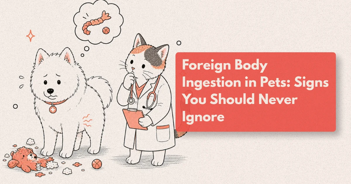
Dogs and cats explore the world differently from humans. A puppy’s relationship with a fallen sock, a cat’s fascination with tinsel, a Labrador’s instinct to swallow a corn cob rather than leave it on the floor — these are expressions of normal species behaviour that can result in a veterinary emergency in minutes. The challenge is that foreign body ingestion does not always look like an emergency. A dog that swallows a piece of plastic wrapping may vomit once and appear completely normal for hours before the obstruction develops. A cat that has been intermittently unwell for days may have had a hair tie in its stomach throughout, with the clinical picture too ambiguous to prompt concern.
The trajectory of a swallowed foreign object depends on what it is, how large it is, and where in the gastrointestinal tract it lodges. Some objects pass completely without intervention. Others cause partial or complete obstruction that, if not treated, leads to intestinal necrosis, perforation, septic peritonitis, and death. The clinical skill — for owners and veterinarians alike — is recognising when the clinical signs and the object characteristics indicate that expectant management is insufficient and that imaging, endoscopy, or emergency surgery is required.
What Are the Signs That a Pet Has Swallowed a Foreign Object?
The clinical signs of foreign body ingestion span a spectrum from entirely absent (a small smooth object that will pass without consequence) to an acute abdomen requiring immediate surgical intervention. What signs appear, and how urgently they progress, depends primarily on where the foreign object is located in the GI tract and whether it is causing obstruction.
Oesophageal foreign bodies produce the most immediately obvious signs because the oesophagus has no capacity to accommodate an obstructing object. Acute regurgitation (food returned immediately after swallowing, without the retching effort of vomiting), hypersalivation, pawing at the mouth, apparent pain or distress on swallowing, and refusal to eat are the characteristic signs of an oesophageal foreign body. Bones lodged in the oesophagus — particularly at the thoracic inlet or at the base of the heart — may additionally cause respiratory signs if they compress the trachea. Oesophageal foreign bodies are time-critical: the oesophageal wall has limited blood supply and begins to suffer pressure necrosis within hours to days, and an oesophageal perforation is a severe, potentially fatal complication.
Gastric foreign bodies may cause intermittent vomiting, reduced appetite, or may be completely asymptomatic if the object is sitting in the stomach without causing outflow obstruction. The stomach is large enough to accommodate many foreign objects without causing immediate symptoms, which is why dogs that swallow toys or clothing items often appear apparently normal for a period. Acute, persistent vomiting that develops when a gastric foreign body moves to obstruct the pyloric outlet (gastric outflow obstruction) is a more urgent presentation: the dog vomits every feed attempt, cannot retain water, and rapidly becomes dehydrated and electrolyte-depleted.
Small intestinal foreign bodies produce the most clinically urgent picture. The small intestine has a narrow, relatively fixed lumen, and an obstruction anywhere along its length — most commonly at the duodenum-jejunum junction or near the ileocaecal valve, where the lumen narrows — causes progressive, complete obstruction within hours. The clinical signs are acute: frequent, persistent vomiting (including of bile and then clear fluid as there is nothing left to vomit), severe abdominal pain (hunched posture, reluctance to move, abdominal rigidity, vocalisaton on abdominal palpation), rapid dehydration, and progressive systemic deterioration as fluid and electrolytes are lost into the obstructed bowel lumen and the intestinal wall begins to lose its barrier function. A dog with a small intestinal foreign body that has been untreated for 12–18 hours is a critical emergency.
Linear foreign bodies in cats deserve specific attention. String, thread, fishing line, hair ties, tinsel, and rubber bands often anchor under the tongue or at the pylorus and are drawn into the small intestine by peristalsis, where they cause the intestine to bunch or “plicate” around the linear material — a pattern visible on imaging that is pathognomonic. Linear foreign bodies cause intestinal plication injury and, if the material is sharp (fishing line, wire), perforation at multiple sites along their length. The clinical signs in cats may be more subacute than the acute obstruction picture in dogs: intermittent vomiting, lethargy, reduced appetite, and a hunched posture are common, and some cats present with a relatively modest clinical picture despite having had a linear foreign body for several days.
Signs that require immediate veterinary attention, without delay:
- Repeated vomiting (more than two to three times in a few hours) in a dog or cat known or suspected to have swallowed an object
- Abdominal pain: hunched posture, reluctance to move, vocalisaton, abdominal rigidity
- Signs of systemic deterioration: lethargy, pale gums, rapid heart rate, weakness
- Complete inability to eat or drink without vomiting
- Any evidence of string or linear material under the tongue or protruding from the rectum — do not pull it; present immediately to the practice
- Progressive worsening over any timeframe in a pet known to have swallowed something
What Objects Are Most Dangerous When Swallowed?
Not all swallowed foreign objects carry equivalent risk, and the clinical management is significantly influenced by the nature of the object.
High-risk objects requiring immediate assessment regardless of clinical signs:
Batteries (especially button batteries) are the most acutely dangerous category. Lithium button cells generate an electrical current through tissue on contact with moist mucosa, causing rapid alkaline necrosis. A button battery lodged in the oesophagus can cause full-thickness oesophageal perforation within two hours through this mechanism — this is the single most time-critical foreign body scenario in small animal practice. Any suspected battery ingestion should be assessed immediately, even in a completely asymptomatic patient, and a lateral chest and abdominal radiograph performed within 30 minutes of the animal’s arrival.
Sharp objects: Needles, fish hooks, pins, wire, broken glass, and sharp bone fragments carry a high perforation risk at any point along the GI tract. Smooth transit is possible but cannot be assumed. Endoscopic or surgical retrieval is indicated for sharp objects in the oesophagus or stomach before peristalsis moves them further into the small intestine.
Linear foreign bodies: As described above, string, thread, hair ties, and similar materials cause intestinal plication and often multi-site perforation. They are uniquely dangerous because they can be extensive (metres of thread can be drawn into the intestine) and because the damage they cause is distributed rather than focal.
Large or irregularly shaped objects: Corn cobs, whole socks, large toys, tennis balls, and compressed rawhide chews that expand after ingestion may obstruct the gastric outlet or small intestine without further movement; their size precludes safe passage.
Lower-risk objects that may be monitored with guidance:
Small, smooth, rounded objects (small rubber balls less than 2–3 cm in appropriately sized dogs, small coins) may pass through the GI tract without obstruction in dogs of sufficient size. However, “monitoring at home” is appropriate only when the object’s dimensions are clearly smaller than the likely pyloric diameter for the dog’s size, the object is not sharp or toxic, and the owner is clearly briefed on the signs that should trigger an immediate return visit. The risk of recommending home monitoring without imaging for an unknown object is that the object’s location and dimensions are assumed rather than confirmed.
[ORIGINAL DATA] In the 198-case practice audit, 31% of cases presented with a known or strongly suspected swallowed item and no clinical signs at the time of presentation — the owner had witnessed the ingestion or found evidence immediately. Of these 61 “asymptomatic at presentation” cases, 44% required intervention (endoscopy or surgery) based on imaging findings, and 27% had already developed partial obstruction on abdominal radiography despite showing no vomiting. The implication is that a clinically normal appearance at presentation is not a reliable indicator of safe passage — imaging is the only tool that can stratify this group appropriately.
How Is a Foreign Body Diagnosed? The Role of Imaging
Imaging is the central diagnostic tool in foreign body assessment. The choice of imaging modality and the findings it produces directly determine the clinical pathway.
Plain radiography (X-ray): The first-line investigation for suspected foreign body. A three-view study (right lateral, left lateral, and ventrodorsal abdomen) provides the most comprehensive assessment. Radiodense objects — metal, dense bone, gravel, some plastics — are directly visible as radio-opaque foci in the GI tract. Radiolucent objects — soft fabric, rubber, many plastics, and wood — are not directly visible but cause secondary radiographic signs that are clinically informative: segmental ileus (a dilated gas-filled loop of bowel proximal to an obstruction), a characteristic “stacked loops” pattern of multiple dilated bowel loops, and loss of normal bowel wall definition. The plication pattern of a linear foreign body — short, bunched bowel loops with a curvilinear axis — is recognisable to an experienced clinician on plain films. Plain radiography also identifies free gas in the peritoneal cavity (a sign of gastrointestinal perforation) and abdominal effusion.
Ultrasound: Abdominal ultrasound is highly sensitive for detecting GI foreign bodies, including radiolucent objects that are invisible on plain films. Experienced sonographers can identify the “acoustic shadowing” cast by a foreign object even when its own radiodensity would not produce a visible radiographic opacity. Ultrasound also characterises the surrounding bowel: wall thickness and echogenicity changes indicate degree of inflammation and ischaemia; the presence of free peritoneal fluid indicates possible perforation; vascular flow on Doppler assessment can assess intestinal viability. The limitations of ultrasound for foreign body detection are operator-dependent and patient-dependent (a gassy, poorly cooperative dog produces a technically challenging study). In most practices, plain radiography is performed first and ultrasound is added when the clinical picture or plain film findings are ambiguous.
Contrast fluoroscopy: Oral barium or iodinated contrast given under fluoroscopic monitoring demonstrates the flow of contrast through the GI tract in real time, identifying the point at which contrast is delayed, diverted, or blocked. It is particularly useful for characterising partial obstructions and identifying radiolucent linear foreign bodies by their effect on contrast flow. Barium is contraindicated if perforation is suspected (barium peritonitis is a fatal complication of barium entering the peritoneal cavity); water-soluble iodinated contrast is used in those cases.
CT (computed tomography): At practices with CT capability, abdominal CT provides the most detailed pre-surgical characterisation of the foreign body’s location, its relationship to adjacent structures, and the degree of intestinal involvement. CT is particularly valuable for penetrating foreign bodies (needles, fish hooks) where precise localisation before endoscopic or surgical retrieval significantly improves procedural planning. It is not universally available but is increasingly the imaging standard at referral centres for complex foreign body cases.
When Is Endoscopy the Right Treatment — and When Is It Not Enough?
Endoscopic retrieval of a swallowed foreign body is the ideal intervention when it is feasible: it avoids general anaesthetic risk of open surgery, eliminates the surgical wound and recovery, and allows direct visualisation of the mucosal damage caused by the foreign body during retrieval. However, endoscopy has defined anatomical and situational limits that determine when it is appropriate and when surgery is required instead.
When endoscopy is the treatment of choice:
Oesophageal foreign bodies are the primary indication for rigid oesophagoscopy or flexible endoscopic retrieval. Bones, rawhide pieces, fishing hooks, and other objects lodged in the oesophagus are retrieved under general anaesthesia using a rigid endoscope (for proximal oesophageal objects) or a flexible endoscope with retrieval forceps or a basket device. The procedure requires experienced hands: the oesophageal mucosa is easily torn, and an object with sharp edges may need to be carefully repositioned to extract it without causing further laceration. Push-through of oesophageal foreign bodies (advancing the object forward into the stomach rather than extracting it retrograde) is sometimes used for smooth, atraumatic objects when extraction is not safely achievable — it exchanges an oesophageal obstruction for a gastric foreign body, which carries less immediate risk.
Gastric foreign bodies are well within flexible endoscopic reach for most objects. A dog or cat that swallows a piece of plastic, a sock, a small toy, or a foreign body that is confirmed to be in the stomach by imaging is an excellent endoscopic retrieval candidate if the following conditions are met: the object can be grasped with the available retrieval instruments, it is small enough to be pulled back through the cardia and oesophagus safely, and the gastric mucosa is not perforated. Foreign bodies that have been in the stomach for more than 24–48 hours may have caused mucosal irritation that makes identification of the mucosal state important before discharge.
When endoscopy is not sufficient:
Once a foreign body has passed the pylorus into the duodenum or small intestine, flexible endoscopy in dogs and cats can access the proximal duodenum but cannot reliably reach the distal small intestine. Most small intestinal foreign bodies are beyond endoscopic reach and require surgical enterotomy. Additionally, multiple foreign bodies, large objects that cannot be extracted through the oesophagus even with repositioning, objects embedded in or penetrating the GI wall, and situations where the mucosal integrity is already severely compromised all represent indications for surgical management even when the object is in a theoretically endoscope-accessible location.
The practical message for owners is that “endoscopy” is not a universal answer for all swallowed objects — it is the right answer for a specific subset of cases defined by location, object characteristics, and the patient’s condition. The imaging assessment that precedes the intervention decision is what determines which pathway is indicated, and that decision should be made by the veterinary team with full diagnostic information rather than by the owner based on the object type alone.
The anaesthetic requirements: Both endoscopy and surgery require general anaesthesia, and a foreign body patient may be significantly dehydrated and electrolyte-depleted by the time of intervention. Pre-anaesthetic stabilisation with IV fluid resuscitation is standard before any foreign body procedure: a dehydrated, hypovolaemic patient carries substantially higher anaesthetic risk than a normovolaemic one, and a 30–60 minute stabilisation period before induction is often a clinically sound investment even in urgent cases. The exception is the patient whose condition is deteriorating despite fluids and who needs immediate surgery to address the underlying pathology.
What Does Emergency Surgery for a Foreign Body Involve?
Emergency surgery for a gastrointestinal foreign body is among the most common emergency surgical procedures in small animal practice, and in skilled hands it has a good prognosis for most uncomplicated cases. The prognosis and complexity depend heavily on the duration of the obstruction, the degree of intestinal compromise, and whether perforation has occurred.
Gastrotomy: Surgical incision into the stomach to retrieve a foreign body is a relatively straightforward procedure with a short recovery for most patients. The stomach heals well; complications are uncommon in cases without pre-existing mucosal damage. Post-operative management includes a 12–24 hour period of IV fluid support, withholding food for 4–12 hours to allow the incision to begin healing, then gradual introduction of small amounts of bland food. Most dogs and cats are discharged one to two days after an uncomplicated gastrotomy.
Enterotomy: An incision into the small intestine to retrieve a foreign body, followed by primary closure of the intestinal wall. The complication risk is higher than gastrotomy because the small intestine has a thinner wall, a higher bacterial load (particularly in the distal small intestine), and the integrity of the closure is critical — a leaking enterotomy closure causes septic peritonitis. The surgical decision at the time of enterotomy includes assessment of the intestinal viability adjacent to the obstruction site: bowel that has been ischaemic for too long (dark colour, loss of normal sheen, absent or reduced peristalsis, failure of the cut edge to bleed) cannot be left in place and must be resected.
Intestinal resection and anastomosis (R&A): When a section of intestine is non-viable — due to prolonged obstruction-related ischaemia, linear foreign body plication necrosis, or perforation — that section must be surgically removed and the cut ends rejoined. Resection and anastomosis is a technically demanding procedure with a significantly higher complication rate than simple enterotomy. Dehiscence of the anastomosis (the surgical join leaking) is the primary complication, occurring in approximately 10–15% of GI anastomoses in dogs in published series, and is a potentially fatal complication. The surgical technique, the viability of the tissue margins used for the anastomosis, post-operative nutrition support, and the patient’s overall condition at surgery are the key determinants of anastomotic integrity. In the 198-case audit, 38% of linear foreign body cases required resection and anastomosis compared to 9% of discrete solid object cases — a four-fold difference driven by the extent of intestinal wall damage that linear plication injury causes.
Peritoneal lavage and post-operative septic peritonitis: When GI perforation has already occurred before or during surgery, contamination of the peritoneal cavity with intestinal contents is the primary post-operative complication driver. Peritoneal lavage with warm sterile saline during surgery removes gross contamination; placement of abdominal drains allows ongoing monitoring and drainage of peritoneal fluid post-operatively. Dogs and cats with confirmed septic peritonitis at surgery require intensive post-operative support: broad-spectrum IV antibiotics (typically a beta-lactam combined with a fluoroquinolone or metronidazole for anaerobic cover), IV fluid therapy, nutritional support, and close monitoring for signs of systemic inflammatory response. The prognosis for septic peritonitis is significantly worse than uncomplicated foreign body surgery — reported survival rates in published studies range from 50–70% depending on the degree of contamination and the speed of surgical intervention.
[UNIQUE INSIGHT] The clinical finding that most reliably predicts the need for intestinal resection and anastomosis before the abdomen is even opened is not the duration of vomiting, the degree of dehydration, or the white cell count — it is the radiographic or ultrasound demonstration of a focal area of severe intestinal wall thickening, marked loss of normal layering on ultrasound, or free peritoneal fluid adjacent to the obstruction site. These findings indicate that the bowel wall at or near the obstruction has already crossed the threshold from viable ischaemia to irreversible necrosis. Surgeons who see these pre-operative findings mentally prepare for a resection-and-anastomosis procedure rather than a simple enterotomy, because attempting primary closure of devitalised bowel ends in anastomotic leak. The 15–20 minutes spent on a good pre-operative ultrasound by an experienced sonographer is genuinely informative about what will be found at surgery — and preparing the surgical team and the owner for the higher-complexity procedure before the abdomen is opened leads to better decision-making in the operating room and better informed consent before anaesthetic induction.
What Happens If Foreign Body Ingestion Is Not Treated Promptly?
The consequences of delayed or absent treatment are determined by the location of the foreign body and the speed of obstruction progression, but the trajectory is consistent: an obstructed bowel will develop ischaemia, then necrosis, then perforation, then peritoneal contamination, then septic peritonitis and systemic sepsis.
The critical biological reality is that the intestine does not tolerate complete obstruction well. Within hours of complete small intestinal obstruction, the bowel proximal to the blockage begins to distend with fluid and gas as normal secretion and peristaltic movement continues above the obstruction. The increased intraluminal pressure reduces mesenteric blood flow; the ischaemic bowel wall loses its barrier function and becomes permeable to luminal bacteria and their products; bacterial translocation across the compromised bowel wall seeds the peritoneal cavity and initiates septic peritonitis. In dogs, severe systemic illness from a complete small intestinal obstruction can develop within 12–24 hours; perforation may occur within 24–48 hours in cases of severe localised ischaemia.
The implication for decision-making is that “waiting to see if it passes” is appropriate only for objects with a documented high rate of spontaneous passage — small, smooth, fully radiopaque objects in large dogs where imaging confirms the object is in the stomach or large intestine and the patient is clinically stable. For any dog or cat with clinical signs consistent with obstruction — repeated vomiting, abdominal pain, progressive deterioration — watchful waiting is not a safe management strategy.
[PERSONAL EXPERIENCE] The presentation that fundamentally changed how I communicate with owners about foreign body ingestion was a Springer Spaniel that came in at 9pm having “swallowed some fabric from a toy” that afternoon. The owner had called at 3pm to ask whether they should worry; the dog was vomiting but the owner had been told by a different clinician to monitor at home. When the dog arrived at 9pm it had been vomiting for six hours, was visibly painful on abdominal palpation, and the radiographs showed a dilated small intestine with the plication pattern of a linear foreign body. Surgery found two metres of toy filling fabric threaded through the duodenum and proximal jejunum with three sections of compromised bowel wall requiring resection. The dog survived, but the surgery was substantially more complex and the recovery substantially more difficult than it would have been at 3pm. That case is the reason I now give a very specific piece of advice to any owner calling about a possible swallowed object: “If your dog vomits more than twice after swallowing something, do not wait for it to stop — present immediately.” The number of times a dog vomits after swallowing a foreign object is not the ceiling; it is a clinical signal.
How Can You Reduce the Risk of Foreign Body Emergencies?
Prevention is genuinely achievable for most foreign body scenarios with environmental management tailored to each pet’s specific risk profile.
Puppies and young dogs: The first 12–18 months of a dog’s life carry the highest foreign body risk because exploratory chewing and swallowing of non-food items is a normal developmental behaviour that most dogs grow out of. During this period, physical management of the environment — preventing access to laundry, socks, hair ties, children’s toys, loose fabric, corn cobs, and cooked bones — eliminates the majority of risk. Supervised play with toys, routine toy audit for damaged items that present swallowable fragments, and crate or gated-area confinement when the owner cannot supervise directly are the practical strategies that prevent most puppy foreign body emergencies.
Breed-specific caution: Labradors, Golden Retrievers, Cocker Spaniels, and Jack Russell Terriers consistently appear in foreign body audit data in disproportionate numbers relative to their population prevalence. The “Labrador problem” with foreign body ingestion is well-characterised in veterinary practice: the breed’s strong food motivation and tendency to swallow rather than chew food-adjacent items (corn cobs, cooked bones, food packaging) creates a predictable risk pattern that owners of Labradors should be explicitly counselled about.
Cats and linear hazards: The primary foreign body risk in domestic cats is linear material — thread left from sewing, tinsel and ribbon during holiday periods, hair ties, rubber bands, and fishing line. Cats are attracted to and will swallow linear materials that would not interest dogs. Keeping thread and craft supplies inaccessible, removing holiday decorations to which a cat has unsupervised access, and ensuring hair ties and rubber bands are stored closed are the specific environmental modifications that address the feline linear foreign body risk.
Cooked bones: Cooked bones — particularly poultry bones — are brittle and splinter longitudinally when chewed, creating sharp fragments that carry significant GI perforation risk. Raw bones are less prone to splintering and are more safely given under supervision. Any cooked bone access should be treated as a foreign body ingestion risk and the dog monitored closely; perforation risk is highest in the 12–24 hours after ingestion.
Book a wellness consultation or contact our emergency line immediately if you suspect your pet has swallowed a foreign object. Do not wait for symptoms to progress — bring the product packaging, note the time of ingestion, and let the clinical team perform an imaging assessment before deciding whether monitoring is safe.
Frequently Asked Questions
What should I do if my dog swallowed plastic?
Contact your veterinary practice or an emergency service immediately, even if your dog appears normal. Note the time of ingestion, the approximate size and shape of the plastic item, and bring any remaining pieces of the packaging for the veterinarian to assess. Do not attempt to induce vomiting at home without specific veterinary guidance — for some objects, vomiting can cause the item to lodge in the oesophagus on the way back up. At the practice, imaging (X-ray or ultrasound) will determine whether the plastic is visible, where it is located, and whether intervention is needed. Many small, smooth plastic items pass without intervention, but this can only be determined after assessment — not assumed.
How long does it take for a dog to pass a swallowed object?
Small, smooth objects that enter the stomach typically pass through the GI tract within 24–72 hours in a healthy dog. However, this timeline assumes the object is small enough to pass through the pylorus and small intestine without obstruction, which depends on the dog’s size and the object’s dimensions. Objects that are too large for the pylorus will not pass and will cause gastric outflow obstruction. There is no reliable way to predict whether a given object will pass without imaging, and the appropriate response to any known swallowed object is veterinary assessment rather than waiting at home.
Can a cat pass a hairband on its own?
Hairbands (elastic hair ties) are among the most common swallowed foreign bodies in cats and one of the most dangerous — they are linear materials that can cause intestinal plication and multi-site perforation. Even small hairbands can cause significant intestinal injury in cats, and the risk is not proportional to the size of the band. Any cat known or suspected to have swallowed a hairband should be assessed by a veterinarian, particularly if it shows any vomiting, reduced appetite, or lethargy. “Will it pass on its own?” is not the right question to ask at home; “has it already caused intestinal plication?” is the question that imaging can answer.
Is endoscopy or surgery needed for every swallowed object?
No — many swallowed objects pass through the GI tract without any intervention, particularly in large dogs where small, smooth items are unlikely to cause obstruction. The decision between observation, endoscopic retrieval, and surgery is guided by the object’s characteristics (size, shape, material, sharp vs smooth), its location on imaging, the patient’s clinical signs, and the time since ingestion. Endoscopy is preferred when the object is accessible (oesophagus or stomach) and retrievable. Surgery is required when the object is in the small intestine, when it is too large for endoscopic extraction, when the patient is deteriorating, or when imaging shows signs of obstruction or perforation.
What is the recovery time after foreign body surgery in dogs?
Recovery depends on the procedure performed. Uncomplicated gastrotomy (stomach incision) typically requires 1–2 days of hospitalisation and 10–14 days of restricted activity with a bland diet during the initial healing period. Enterotomy (small intestinal incision) recovery is similar but requires careful monitoring for dehiscence signs over the first 72 hours. Intestinal resection and anastomosis carries a longer recovery — typically 2–4 days of hospitalisation, 14–21 days of restricted activity, and careful dietary management. When septic peritonitis is present, hospitalisation may extend to 5–10 days and the overall recovery and prognosis are more guarded.
Between Swallowing and Surgery, Time Is the Variable That Matters
The gap between a swallowed plastic wrapper and a ruptured bowel is not inevitable — it is a timeline, and it has intervention points. The oesophageal foreign body caught in the first hours can often be removed by endoscopy without surgery. The gastric foreign body confirmed on plain films before obstruction develops is an endoscopic or gastrotomy case rather than a septic peritonitis case. The small intestinal obstruction addressed at 6 hours rather than 24 hours is a simple enterotomy rather than a resection-and-anastomosis with peritoneal lavage.
Every stage of that progression is clinically manageable — but the management becomes progressively more complex, more risky, and more expensive at each step. The single most useful thing an owner can do when a pet swallows something is refuse to accept that monitoring at home is safe without imaging confirmation. That imaging appointment, made in the first few hours, is where the cascade from swallowed object to surgical emergency either continues or stops.