CCL Tear in Dogs: Causes, Diagnosis, and Recovery
CCL rupture is the most common orthopedic injury in dogs. Learn why ligaments fail, how it's diagnosed, and what TPLO surgery and recovery really involve.
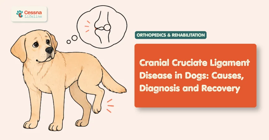
Cranial cruciate ligament (CCL) rupture is the most common orthopedic condition in dogs and the leading reason for hind limb surgery in small animal practice (Fossum, Small Animal Surgery). Hundreds of thousands of dogs undergo surgical repair in the United States every year, with estimates placing the annual case load above 600,000 and total associated veterinary costs exceeding one billion dollars (Wilke et al., 2005). For many families, a CCL injury is the most significant veterinary event their dog will experience.
Understanding why the ligament fails, what the diagnosis involves, what surgical options mean, and what recovery actually looks like removes the uncertainty that makes these situations feel more overwhelming than they need to be. dog limping evaluation
What to Know CCL rupture is the most common orthopedic injury in dogs and affects all breeds, though large and giant breeds carry disproportionate risk. It is almost never a single traumatic event; it is a progressive degenerative process. Surgery is the standard of care for dogs over 15 kilograms. TPLO is the most widely performed repair technique, with good-to-excellent long-term outcomes in approximately 85 to 93% of dogs (Fossum; Bergh & Peirone, 2012).
The Knee Joint That Fails Without Warning
The canine stifle joint (the anatomical equivalent of the human knee) is stabilized by four ligaments: the cranial cruciate, the caudal cruciate, and two collateral ligaments. The cranial cruciate ligament (CCL), equivalent to the anterior cruciate ligament (ACL) in humans, runs diagonally through the center of the joint and prevents the tibia from sliding forward relative to the femur during weight-bearing. When it fails, the joint becomes unstable, causing pain and progressive cartilage and meniscal damage with every step.
The critical difference between canine CCL disease and human ACL tears is the mechanism. Human ACL ruptures are overwhelmingly traumatic: a single pivoting event, a collision, a landing from a jump. In dogs, CCL rupture is almost always the endpoint of a chronic degenerative process. The ligament undergoes progressive structural degradation over months to years, with collagen fiber fragmentation, decreased vascular supply, and inflammatory cell infiltration occurring well before the ligament finally tears (Hayashi et al., 2003). The moment of apparent "sudden" lameness is the final structural failure of a ligament that was already compromised.
This distinction matters enormously for owner understanding. When a dog goes suddenly lame after playing fetch or jumping off the couch, the natural interpretation is "my dog just injured his knee." The more accurate interpretation is "my dog's ligament finally failed after months of degeneration, and this activity was the event that crossed the threshold." The activity did not cause the injury; the ligament was already failing. This reframing matters because it explains why dogs are often lame on a limb before any obvious injury event, why resting "to let it heal" does not resolve the problem, and why the contralateral limb faces the same degenerative process independently.
Why Do Dogs Tear Their Cruciate Ligaments?
The etiology of canine CCL disease is multifactorial, involving conformational, genetic, inflammatory, and body condition factors. No single cause explains all cases, and the interplay between these factors is still being characterized in the research literature.
Tibial plateau angle (TPA): The slope of the tibial plateau (the top surface of the tibia) is a major biomechanical contributor. A steeper tibial plateau creates greater cranial tibial thrust during weight-bearing, placing more tensile load on the CCL with every step. Dogs with high tibial plateau angles, particularly in breeds with straight rear legs and steeply angled tibias, experience higher CCL loading across their lifetime. This is the biomechanical basis for the TPLO procedure, which reduces the tibial plateau slope to eliminate shear force rather than replacing the ligament.
Body weight and obesity: Overweight and obese dogs are at substantially elevated risk for CCL disease. Excess body weight increases joint load and accelerates ligament fiber degeneration. Dogs with a body condition score above 5 out of 9 carry measurably higher CCL rupture rates than lean dogs (Faries et al., 2021). Obesity also increases the risk of contralateral limb rupture after the first tear. dog weight management
Breed predisposition: CCL disease is strongly influenced by genetics, with certain breeds carrying dramatically elevated lifetime risk. Rottweilers, Labrador Retrievers, Newfoundlands, Staffordshire Bull Terriers, and West Highland White Terriers are among the most consistently cited high-risk breeds (Witsberger et al., 2008). Interestingly, Greyhounds and Dachshunds show very low CCL disease rates, likely due to conformational differences in tibial plateau angle and joint mechanics.
Age and sex: Middle-aged dogs (3 to 7 years) are most commonly affected, though CCL disease can occur at any age. Neutered males and spayed females show higher rates than intact animals in multiple studies, suggesting a hormonal component to ligament integrity. Early neutering before skeletal maturity has been associated with elevated CCL rupture risk in large breeds in several independent analyses.
Citation Capsule: Rottweilers carry an approximately 5-fold elevated lifetime risk of CCL rupture compared to mixed-breed dogs, while Greyhounds and Dachshunds appear at the low end of breed-specific incidence, likely due to conformational differences in tibial plateau angle that reduce cranial tibial thrust during weight-bearing (Witsberger et al., 2008). Neutered males and spayed females show higher CCL rupture rates than intact animals across multiple independent analyses, with early neutering before skeletal maturity associated with additional risk elevation in large breeds.
What Does a CCL Tear Look Like in a Dog?
The most common presentation is a dog that goes suddenly lame on a hind limb after normal activity: playing in the yard, jumping off furniture, or coming in from a walk. The dog typically holds the leg up or toe-touches briefly, and many owners note that the dog is not crying or yelping, which creates a confusing picture. The absence of vocalization does not reflect a minor injury; it reflects the dog's stoic pain tolerance and the nature of the ligament injury itself. dog limping urgency
In acute complete tears, the dog is typically non-weight-bearing or minimally weight-bearing on the affected limb within minutes to hours of the event. In partial tears, the dog may be moderately lame, may improve over a few days with rest (only to relapse with activity), and may cycle through periods of apparent improvement followed by recurrence. This cycling pattern, lameness that seems to respond to rest but returns with exertion, is highly characteristic of partial CCL disease that has not yet progressed to complete rupture.
On physical examination, key findings include joint effusion (fluid accumulation causing the stifle to feel thicker than normal), pain on full stifle extension, and a positive cranial drawer test or tibial compression test. The cranial drawer test demonstrates abnormal forward sliding of the tibia relative to the femur when the examiner stabilizes the femur and applies cranial pressure to the tibia; this movement should not be present in a healthy stifle. In very muscular dogs or in acute injuries with significant muscle spasm, the drawer test may require sedation to elicit clearly.
What orthopedic surgeons observe consistently: The overwhelming majority of owners who present with a dog that went suddenly lame "playing fetch" or "jumping off the couch" describe the event as an injury, and express concern about what the dog did to its knee. The conversation that almost always follows is an explanation that this was not an injury in the traumatic sense: the ligament was already degenerating, and the activity was incidental to the final failure. Many of these dogs were intermittently lame in the weeks before the acute event, or showed subtle reluctance on stairs or getting up after rest, but those signs were attributed to "overdoing it" or normal stiffness. Owners almost universally say, in retrospect, "now that you mention it, yes."
Citation Capsule: Partial CCL tears represent an intermediate disease stage where the dog may cycle through lameness and apparent recovery; studies indicate that approximately 50 to 60% of partial tears progress to complete rupture without surgical intervention, and dogs with complete tears lose functional meniscal tissue in 50 to 75% of cases when the unstable joint has been loaded for weeks or months before diagnosis (Cook, 2010). Meniscal damage at time of surgery is an independent predictor of longer recovery and reduced long-term outcome quality.
How Do Vets Diagnose a Cruciate Ligament Injury?
Diagnosis begins with the physical examination. A veterinarian who identifies joint effusion, pain on extension, and a positive cranial drawer or tibial compression test has sufficient grounds for a CCL diagnosis without imaging in most cases. However, radiographs are standard practice for several reasons: they rule out concurrent bone pathology (fracture, tumor), assess the degree of osteoarthritis already present, and measure the tibial plateau angle if TPLO is being considered.
Radiographic signs of CCL disease include joint effusion (visible as a soft tissue density displacing the infrapatellar fat pad), periarticular osteophyte formation (indicating chronic instability), and in some cases cranial displacement of the tibia relative to the femur. Radiographs do not visualize soft tissue structures directly; they confirm the orthopedic context around a clinical diagnosis.
MRI and CT are increasingly used in referral orthopedic settings, particularly when partial tears are suspected or when concurrent pathology (meniscal tears, osteochondrosis) needs to be characterized before surgical planning. Arthroscopy, direct visualization of the joint interior with a small camera, is both diagnostic and therapeutic: it allows assessment of the menisci, debridement of damaged ligament tissue, and meniscal release or partial meniscectomy during the same procedure.
The menisci are C-shaped fibrocartilage structures that cushion the joint. The medial meniscus is particularly vulnerable in CCL-deficient stifles because it becomes partially load-bearing in an unstable joint. Meniscal tears at the time of CCL repair are found in approximately 50 to 75% of chronic complete tears, and addressing them during surgery is essential for optimal recovery.
Citation Capsule: Meniscal pathology is present in approximately 50 to 75% of dogs with chronic complete CCL ruptures at the time of surgical exploration, and concurrent meniscal tear is an independent predictor of reduced long-term functional outcome when not addressed (Cook, 2010). Arthroscopic assessment of the stifle at the time of surgical repair allows concurrent meniscal evaluation and treatment in a single anaesthetic event, which is increasingly the standard approach at referral centers.
Which Surgery Is Right: TPLO, TTA, or Lateral Suture?
Three surgical techniques account for the vast majority of CCL repairs in dogs. Each approaches the mechanical problem of cranial tibial instability differently.
CCL Surgery Comparison: TPLO, TTA, and Lateral Suture
| Procedure | Mechanism | Best For | Return to Function | Major Complication Rate |
|---|---|---|---|---|
| TPLO (Tibial Plateau Leveling Osteotomy) | Cuts and rotates tibial plateau to reduce slope, eliminating cranial tibial thrust | Dogs over 15 kg; active dogs; high tibial plateau angle | 12 to 16 weeks | Approximately 5 to 8% |
| TTA (Tibial Tuberosity Advancement) | Advances the tibial tuberosity to change patellar ligament angle and neutralize tibial thrust | Medium to large dogs; alternative geometry indication | 12 to 16 weeks | Approximately 5 to 10% |
| Lateral suture (extracapsular repair) | Synthetic band placed outside the joint mimics CCL function during scar tissue formation | Dogs under 15 kg; older or less active large dogs | 8 to 12 weeks | 10 to 15% long-term |
TPLO (Tibial Plateau Leveling Osteotomy) has become the dominant repair technique in dogs over 15 kilograms and is considered the gold standard in most referral orthopedic centers. Rather than replacing the ligament, TPLO changes the joint geometry by cutting the tibial plateau, rotating it to reduce its slope, and securing it with a bone plate. The result is a joint that is mechanically stable during weight-bearing without relying on a new ligament structure. Published outcomes show good-to-excellent long-term function in approximately 85 to 93% of TPLO patients (Bergh & Peirone, 2012). Most dogs begin partial weight-bearing within days of surgery and are functionally sound at 12 to 16 weeks with appropriate rehabilitation.
TTA (Tibial Tuberosity Advancement) achieves joint stability through a different geometric principle: by advancing the tibial tuberosity anteriorly, it changes the angle of the patellar ligament relative to the tibial plateau such that cranial tibial thrust is neutralized during normal activity. Outcomes are broadly similar to TPLO, and the choice between them is often based on individual joint geometry, surgeon preference, and patient factors.
Lateral suture (extracapsular repair or fabello-tibial suture) places a strong synthetic material outside the joint to provide temporary stabilization while periarticular scar tissue forms. It is the most technically straightforward and least expensive option, and it remains the procedure of choice in dogs under 15 kilograms, where the biomechanical demands on the repair are lower. In large dogs, lateral suture has lower long-term success rates than TPLO or TTA: studies report good-to-excellent outcomes in approximately 50 to 70% of large breed dogs compared to 85 to 93% with TPLO (Fossum).
Citation Capsule: TPLO produces good-to-excellent long-term functional outcomes in approximately 85 to 93% of operated dogs across published case series, with the outcome advantage over lateral suture most pronounced in dogs over 15 kilograms, where lateral suture success rates drop to approximately 50 to 70% at long-term follow-up compared to 85 to 93% with TPLO (Bergh & Peirone, 2012; Fossum, Small Animal Surgery). This size-stratified outcome difference is the primary evidence basis for recommending osteotomy-based techniques over extracapsular repair in large and giant breed dogs.
What Recovery from CCL Surgery Actually Looks Like
CCL recovery is a structured, phased process that cannot be rushed without compromising outcomes. The timeline for TPLO and TTA is similar: most dogs are using the leg for light weight-bearing within a few days of surgery, but return to normal activity takes 12 to 16 weeks, and full muscular recovery extends beyond that.
Phase 1 (Weeks 0 to 4): Strict rest and wound management. The dog is restricted to leash walks only, 5 to 10 minutes three to four times daily. No running, jumping, stairs (where avoidable), or off-leash activity. Ice packing the surgical site for 10 to 15 minutes three to four times daily reduces swelling. Passive range-of-motion exercises, gently flexing and extending the operated joint while the dog is lying down, maintain joint mobility and prevent scar tissue contracture during this phase.
Phase 2 (Weeks 4 to 8): Controlled exercise increase. Leash walks progressively lengthen. Many rehabilitation protocols introduce hydrotherapy (underwater treadmill or swimming) during this phase, which allows the dog to exercise limb muscles without full weight-bearing impact. Physiotherapy assessment and a structured rehabilitation plan, rather than owner-managed home exercise alone, consistently produces better muscular outcomes.
Phase 3 (Weeks 8 to 16): Progressive return to function. Leash walks extend to 20 to 30 minutes, and the surgical site is radiographically assessed to confirm bone healing in TPLO and TTA patients. Off-leash activity and full running return only after radiographic confirmation of healing and clinical assessment of weight-bearing symmetry. dog rehabilitation therapy
Non-surgical management is not a reasonable alternative for dogs over 15 kilograms. Without surgery, approximately 80 to 90% of large breed dogs with complete CCL tears do not achieve acceptable long-term function; ongoing joint instability accelerates cartilage destruction and osteoarthritis progression with every step (Johnson et al., 2016). In dogs under 15 kilograms, conservative management with strict rest, weight management, and physiotherapy achieves acceptable functional outcomes in approximately 80 to 85% of cases, making it a genuine clinical option for the smallest patients.
Will the Other Knee Eventually Tear Too?
One of the most important but least-communicated statistics in canine orthopedics is the contralateral limb rupture rate. Within 1 to 2 years of a first CCL tear, 30 to 60% of dogs will rupture the CCL in the opposite stifle (Buote et al., 2009). This is not because the surgery damaged the other leg; it is because the same underlying degenerative process that weakened the first CCL is operating identically in the contralateral ligament. The biology does not respect surgical repair.
Knowing this statistic changes how owners approach the recovery period and the months that follow. The contralateral leg should be monitored actively. Weight management is particularly important because obesity increases the risk of bilateral disease. Joint supplements (omega-3 fatty acids, glucosamine, chondroitin) have limited evidence for preventing CCL rupture, but supporting joint health and minimizing loading through weight control is the most modifiable risk factor for the second leg. joint supplements dogs
Owners who notice intermittent hindlimb lameness on the opposite side during the recovery period of the first surgery should report it promptly. A partial CCL tear in the contralateral limb during recovery from the first surgery is a known complication of the recovery period itself, because the dog overloads the good leg while protecting the operated one.
Citation Capsule: Contralateral stifle CCL rupture occurs in approximately 30 to 60% of dogs within 12 to 24 months of an index CCL rupture, driven by the same underlying degenerative ligament disease rather than mechanical overload from the initial surgery (Buote et al., 2009). Weight management, because obesity is the most modifiable risk factor for both primary and contralateral CCL disease, is the single most actionable recommendation for reducing second-leg rupture risk during and after recovery from CCL surgery.
Frequently Asked Questions
Can a dog walk on a torn cruciate ligament?
Many dogs with complete CCL tears continue to toe-touch or briefly weight-bear on the affected leg, particularly in the first hours after rupture. Dogs with partial tears may walk almost normally and only show obvious lameness after exercise. The ability to bear some weight does not indicate a minor injury; it reflects the dog's pain tolerance and the variable degree of joint instability. A dog limping on a hind leg without crying should be evaluated within one to two days, not observed at home for a week.
How do I know if my dog has a CCL tear or something else?
Physical examination by a veterinarian is required for diagnosis. The cranial drawer test and tibial compression test are specific for CCL instability and cannot be replicated by owners at home. Other causes of hind limb lameness in dogs include hip dysplasia, luxating patella, muscle strains, and less commonly bone tumors or nerve root disease. Radiographs help distinguish between these differentially. A positive drawer sign combined with joint effusion on palpation is highly specific for CCL disease.
Is TPLO surgery worth it?
For dogs over 15 kilograms with complete CCL tears, TPLO is the most evidence-supported treatment option and produces good-to-excellent long-term function in approximately 85 to 93% of cases. Non-surgical management in this size group is associated with progressive joint deterioration and poor long-term function in 80 to 90% of cases. The investment in TPLO surgery and recovery is substantial, but the alternative for most large breed dogs is not stability with rest; it is progressive arthritis, chronic pain, and functional decline. TPLO cost recovery
How long does CCL surgery recovery take?
For TPLO and TTA, most dogs are partially weight-bearing within a few days. Leash-only restriction applies for the first 8 weeks. Return to off-leash activity and full exercise typically occurs at 12 to 16 weeks following radiographic confirmation of bone healing. Muscle mass recovery and full athletic function may take 4 to 6 months. Lateral suture recovery is approximately 2 to 4 weeks faster but follows a similar phased structure.
What happens if I don't treat a CCL tear in my dog?
In dogs over 15 kilograms, untreated complete CCL rupture leads to progressive joint instability, accelerated cartilage destruction, meniscal tearing, and severe osteoarthritis. Most large breed dogs with untreated CCL disease develop chronic pain and significant functional impairment. In dogs under 15 kilograms, conservative management with strict rest and rehabilitation can achieve acceptable outcomes in approximately 80 to 85% of cases, making it a legitimate option for the smallest patients when surgery is not feasible.
The Limp That Cannot Wait
A dog that goes suddenly lame on a hind leg without crying often gets a day or two of "rest and see if it improves" at home. For some mild muscle strains, that is appropriate. For CCL tears, it is not: the joint continues to sustain meniscal and cartilage damage with every step on an unstable stifle, and the window for the cleanest surgical repair narrows with delay.
The appropriate response to a dog that is suddenly non-weight-bearing or severely lame on a hind leg is a veterinary evaluation within 24 to 48 hours. Diagnosis is usually straightforward, the treatment pathway is well-established, and the outcomes with appropriate surgical repair are genuinely good. The limp that looks like it might resolve on its own almost never does.