Fractures in Pets: When Immediate Care Matters
A broken leg in a dog or cat is a veterinary emergency. Learn the signs of fractures in pets, how they are classified and repaired, and what first aid and recovery involve.
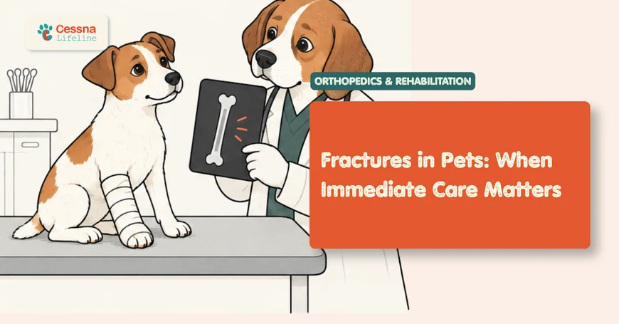
A fracture is one of the clearest orthopedic emergencies in veterinary medicine. Unlike many conditions that can be monitored at home or seen the following week, a fractured bone in a dog or cat requires same-day evaluation. The reason is not merely the fracture itself but what accompanies it: soft tissue damage, vascular compromise, nerve injury, open contamination, and the pain burden that untreated fractures impose with every small movement. Speed in reaching veterinary care directly affects both the treatment options available and the final functional outcome. dog hit by car emergency guide
What to Know Approximately 70% of fractures in dogs result from vehicular trauma. A fractured limb produces non-weight-bearing lameness, visible deformity, and swelling, though cats frequently suppress pain expression and may appear quieter than expected. All fractures require same-day veterinary assessment. Open fractures, where bone is visible through the skin, are contaminated emergencies with a treatment window of 6 to 8 hours before infection is established (Beale, 2004).
The Break That Cannot Wait
Bone fractures in pets occur most commonly from high-energy trauma: vehicular accidents, falls from height, being stepped on (particularly in small breeds and puppies), or dog fights. In older animals, pathological fractures can occur from minimal trauma at sites weakened by bone tumors, the most important of which is osteosarcoma. Understanding that most fractures are traumatic events helps set the right expectations: a fractured pet has often sustained forces capable of damaging not just bone but adjacent vessels, nerves, and organs, and those secondary injuries require as much attention as the fracture itself.
The femur is the most commonly fractured long bone in dogs, accounting for approximately 35 to 40% of all long bone fractures, followed by the radius and ulna, the tibia, and the humerus (Denny and Butterworth, 2000). Pelvic fractures are also common after vehicular trauma, often involving multiple fracture sites simultaneously. In cats, high-rise falls produce a distinctive pattern called "high-rise syndrome" involving facial, thoracic, and limb injuries in combination.
The overarching rule is simple: any pet that has experienced significant trauma and is non-weight-bearing on a limb, or has a visibly abnormal limb position, needs to be seen by a veterinarian the same day, not observed at home overnight.
What Are the Signs of a Fracture in a Dog or Cat?
Recognizing a fracture from the outside involves a specific constellation of findings. Not all are present in every case, but their combination is strongly predictive. pain recognition in cats
The cardinal signs of a fracture in dogs and cats:
- Non-weight-bearing lameness on the affected limb: complete refusal to put the limb down, or brief toe-touching at most
- Visible deformity: the limb at an abnormal angle, shortened, or rotated compared to the opposite side
- Localized swelling: soft tissue swelling developing over the fracture site within minutes to hours
- Crepitus: a grating or crackling sensation if the limb is moved (do not test for this purposely; it causes pain and can worsen soft tissue damage)
- Pain on gentle palpation near the injury site
- Open wound: visible bone end or a puncture wound near the limb in open (compound) fractures
Important distinctions in cats: Cats are profoundly effective at suppressing pain expression. A cat with a fractured limb may not vocalize, may not struggle, and may sit quietly in one position. This stillness is not a sign of being fine; it is a pain behavior. A cat that is immobile and tense following trauma, especially one that flinches when touched, is in significant pain and requires evaluation. pain recognition in cats
Pelvic fractures deserve special mention. A dog or cat with multiple pelvic fractures from vehicular trauma may be able to move all four limbs but drag the hindquarters, show extreme pain when the pelvis is gently compressed laterally, or be unable to urinate or defecate normally if the pelvic canal is narrowed. Urethral and bladder trauma are common concurrent injuries in pelvic fractures.
Citation Capsule: Approximately 70% of fractures in dogs are caused by vehicular trauma, with the femur, radius/ulna, and tibia representing the most commonly affected long bones; a significant proportion of traumatically fractured animals have concurrent thoracic or abdominal injuries that may not be immediately apparent from external examination (Denny and Butterworth, 2000). Initial triage of a fractured pet must therefore include evaluation of respiratory pattern, abdominal pain, and cardiovascular status before focusing exclusively on the limb.
How Do Vets Classify Fractures and Why Does It Matter?
Fracture classification is not academic; it directly determines treatment selection, prognosis, and urgency. Veterinarians use several overlapping classification systems simultaneously when evaluating a fractured bone.
Open versus closed: A closed fracture has intact skin over the fracture site. An open (compound) fracture has a break in the skin communicating with the fracture, whether from the bone end penetrating out or an external wound penetrating in. Open fractures are contaminated by definition and carry substantially higher infection risk. The critical contamination window is 6 to 8 hours: bacteria introduced at the time of injury proliferate to clinically significant levels within that timeframe, after which infection risk escalates sharply and treatment complexity increases (Beale, 2004). Open fractures are treated as emergencies requiring surgical debridement and stabilization, not fractures that can wait until morning.
Fracture configuration:
- Transverse: bone breaks perpendicular to its long axis; relatively stable with fixation
- Oblique: angled break; tends to collapse under load without rigid fixation
- Spiral: rotational force fracture; suggests torsional mechanism
- Comminuted: three or more fragments; greater surgical complexity, longer healing time
- Greenstick: incomplete fracture through cortex; more common in immature animals with flexible bone
Displacement: Whether and how far the fracture ends have shifted from their normal alignment. Non-displaced fractures may heal with conservative management; displaced fractures generally require surgical reduction.
Growth plate (Salter-Harris) classification: In immature animals, the growth plate (physis) is the weakest link in bone, weaker than both ligaments and the adjacent bone. Growth plate injuries are classified on the Salter-Harris system (Types I through V):
| Salter-Harris Type | Description | Prognosis |
|---|---|---|
| Type I | Separation through physis only | Excellent; physis usually intact |
| Type II | Fracture through physis + metaphysis | Good; most common type |
| Type III | Fracture through physis + epiphysis | Fair; involves joint surface |
| Type IV | Fracture through physis + metaphysis + epiphysis | Fair; requires anatomic reduction |
| Type V | Crush injury to physis (compression) | Poor; growth arrest common |
Type V Salter-Harris fractures are the most clinically dangerous growth plate injuries, and they are consistently the most underdiagnosed. Unlike Types I through IV, a Type V injury is a compressive crush of the growth plate rather than a shear or bending fracture. Radiographs at the time of injury can appear nearly normal: no obvious fracture line, minimal displacement. Owners and clinicians may be reassured. But the growth plate cells have been destroyed. Weeks to months later, the affected bone stops growing while the opposite paired bone continues, producing progressive angular limb deformity and shortening. If a puppy has sustained significant compressive trauma to a limb that seems radiographically unremarkable, close radiographic follow-up at 2 and 6 weeks is warranted to detect early growth arrest before the deformity becomes structural.
Fracture First Aid: What to Do Before You Reach the Clinic
Appropriate first aid in the period between the fracture and veterinary care reduces pain, prevents additional soft tissue damage, and reduces contamination in open fractures. Inappropriate first aid, particularly attempting to splint a fracture at home, can cause additional injury.
What to do:
Contain and calm the animal. An injured pet in pain may bite even a trusted owner. Use a towel or blanket to gently wrap and lift the animal, avoiding putting pressure on the fractured limb. For large dogs, a flat board, blanket, or makeshift stretcher can be used to transport without requiring the dog to bear weight. orthopedic surgery in dogs explained
Minimize handling of the fractured limb. Movement at the fracture site causes pain and can worsen soft tissue injury, lacerate vessels, or convert a closed fracture to an open one if a fragment shifts.
For open fractures: cover the wound with the cleanest material available (sterile gauze if you have it, a clean cloth if not). Do not attempt to push the bone back in. Do not pour disinfectants into the wound. The goal is to reduce the bacterial inoculum on the wound surface until surgical debridement can be performed.
Do not administer human pain medications. NSAIDs such as ibuprofen and naproxen are toxic to dogs and cats. Acetaminophen is acutely toxic to cats. Call the veterinary clinic in transit; they can advise on whether any veterinary pain relief you have at home is appropriate.
What not to do:
Do not attempt to splint the fracture at home with rigid materials. An improperly applied splint can compress swelling, compromise blood supply to the limb, and cause pressure necrosis within hours. A padded wrap (such as a soft towel) for transport is acceptable; a rigid splint is not.
Citation Capsule: Open fractures are considered contaminated at the moment of injury; bacterial proliferation to clinically significant levels occurs within 6 to 8 hours, after which infection rates and treatment complexity increase substantially (Beale, 2004). First aid for open fractures should focus on covering the wound with a clean sterile dressing and transporting immediately rather than attempting wound irrigation or manipulation that delays arrival at the clinic.
Which Repair Options Are Available and How Are They Chosen?
Fracture repair in veterinary medicine encompasses a range of surgical and conservative strategies. The choice depends on fracture type, location, the size and age of the animal, the owner's capacity for postoperative management, and cost.
Conservative management (casting and splinting): Appropriate for a narrow set of fractures: incomplete (greenstick) fractures, minimally displaced fractures below the elbow or stifle in small breeds, some growth plate separations, and young animals whose rapid healing biology can tolerate controlled external support. Conservative management requires strict activity restriction, owner compliance with bandage checks, and typically more radiographic monitoring than surgical repair. Complications include pressure sores, cast-associated swelling, and delayed or non-union if the fracture is not adequately stable.
Intramedullary (IM) pinning: A metal pin placed down the hollow medullary canal of the bone to align the fragments. Provides axial alignment but limited rotational and bending stability; often combined with cerclage wire or external fixation to address these limitations. Most useful for simple transverse and short oblique fractures in the femur and humerus of appropriately sized patients.
Bone plate and screw fixation: The gold standard for most long bone fractures in dogs. A metal plate is applied to the cortical surface of the bone and secured with cortical screws that engage both cortices. Provides rigid fixation against axial, bending, and rotational forces, allowing early controlled limb use in most patients. Outcomes consistently exceed 85 to 90% good-to-excellent in appropriately selected cases (Piermattei et al., 2006). orthopedic surgery dogs
External skeletal fixation (ESF): Metal pins are driven through the skin into the bone above and below the fracture, then connected by an external frame. The bone stabilizes while the frame remains outside the body. Particularly valuable for open fractures where bone plating would introduce an implant into a contaminated field, or for highly comminuted fractures. Requires daily pin tract cleaning and careful owner management.
Interlocking nails: A variation on IM pinning with additional locking screws that resist rotation and bending, providing greater stability than a plain IM pin. Used primarily in large-breed femoral and tibial fractures.
Citation Capsule: Bone plate fixation achieves good-to-excellent functional outcomes in approximately 85 to 90% of appropriately selected canine long bone fracture cases, representing the most consistently reliable single surgical technique for mid-diaphyseal fractures of the femur, tibia, radius/ulna, and humerus (Piermattei et al., 2006). The advantage of rigid plate fixation, compared to IM pinning, is resistance to all force vectors simultaneously, which allows earlier controlled weight-bearing and reduces the biological healing time by minimizing micromotion at the fracture gap.
What Does Fracture Recovery Actually Look Like?
Fracture recovery is not a linear process. Bone healing progresses through four overlapping biological phases: hematoma formation (days 1 to 5), soft callus development (weeks 1 to 3), hard callus mineralization (weeks 3 to 12), and remodeling (months to years). What an owner observes at home does not always align with what the radiograph shows, and vice versa.
Activity restriction is the most critical owner-managed variable. The single most common cause of fracture repair failure is inadequate confinement during the healing period. A dog that jumps off a sofa six weeks after femoral plate repair may re-fracture through the healing callus or bend an implant before the bone has reached adequate strength. Strict crate rest or room confinement, leash-only outdoor access for toileting, and prohibition of stairs and jumping are the core early-recovery requirements for most fractures. post-surgery rehabilitation dogs
Radiographic re-evaluation is performed at intervals of typically 6 to 8 weeks to assess callus formation and bone bridging. The fracture is considered healed when radiographic evidence of bone bridging across the fracture line is present; this does not always correlate with return to full activity. In young animals, healing progresses much faster than in older ones: a 10-week-old puppy may show radiographic healing within 3 to 4 weeks; an 8-year-old dog may require 10 to 16 weeks.
What emergency clinicians observe consistently: The single most predictable pattern in fracture aftercare is the owner who reports at the 6-week recheck that the dog "seemed fine at 3 weeks so we let him play a bit." Bone callus is radiographically visible at 3 to 4 weeks but is not load-bearing. It has the consistency of hard rubber, not cortical bone. Allowing unrestricted activity before bridging callus has mineralized does not mean the pet feels pain; dogs with hardware in place often feel dramatically better before the bone is healed, because the implant is bearing the load. The implant is not a replacement for bone; it is a temporary scaffold. Protect the scaffold until the bone takes over.
Physiotherapy and controlled rehabilitation exercise become important in the intermediate recovery phase (weeks 4 to 12), targeting the muscle atrophy and proprioceptive deficits that accumulate during the period of enforced rest. A dog that was crate-rested for 8 weeks has significant muscle loss in the affected limb that does not spontaneously resolve with free exercise. Structured rehabilitation, including range-of-motion exercises, walking on varied surfaces, and hydrotherapy, consistently improves functional return timelines. post-surgery rehabilitation dogs
Citation Capsule: Structured physiotherapy and hydrotherapy initiated in the intermediate fracture recovery phase (weeks 4 to 12 post-repair) significantly improves limb muscle mass recovery and functional limb use compared to unstructured rest-to-activity transition; the muscle atrophy accumulated during strict confinement does not reverse spontaneously with free exercise alone and requires active rehabilitation to restore periarticular support (Millis and Levine, 2014). Rehabilitation also accelerates proprioceptive recovery, which reduces re-injury risk during the final return-to-function phase.
Does Age, Size, or Location Change the Outcome?
Yes, substantially, and in ways that are not always intuitive.
The toy breed radius/ulna problem: Distal radius and ulna fractures in toy and miniature breeds (Yorkshire Terriers, Chihuahuas, Pomeranians, Miniature Poodles, and similar) have a uniquely high complication rate that is fundamentally misunderstood as a size problem. It is not. It is a vascular anatomy problem. The distal radial metaphysis in toy breeds has markedly poor periosteal blood supply, combined with a thin cortex and minimal surrounding soft tissue coverage, relative to medium and large breeds. This combination produces delayed union and nonunion rates of 40 to 60% when these fractures are managed conservatively with casting alone, even when the fracture is minimally displaced and the cast is well-applied (Welch et al., 1997). Surgical fixation with small bone plates or external fixators is now recommended as the primary treatment for most distal radius fractures in toy breeds, not as a last resort. Owners should be counseled on this upfront rather than after a failed conservative attempt. toy breed health concerns
Age and growth plate vulnerability: In animals under 12 months of age, the growth plate (physis) is weaker than surrounding structures. A force that would sprain a ligament in an adult will fracture through the growth plate in a puppy. Growth plate fractures carry specific prognoses based on Salter-Harris type and the specific physis involved. The distal femoral physis fracture (Salter-Harris Type I or II), one of the most common growth plate injuries in cats and young dogs, carries a good prognosis with early surgical repair but a poor prognosis if delayed or managed conservatively. growth plate injuries puppies
Cats and open fractures: Cats are overrepresented in high-rise falls and vehicular accidents that produce complex, comminuted, and often open fractures. Feline bone is more brittle relative to body size than canine bone, and cat fractures more often involve multiple bones simultaneously. Cats also tolerate certain implants differently; cats show higher rates of implant loosening with external fixators than dogs, and their smaller bone size limits plate and screw options. Despite this, outcomes after surgical repair in cats are generally good when performed by an experienced surgeon with appropriately scaled implants.
Citation Capsule: Toy breed distal radius/ulna fractures managed conservatively with casting have delayed union or nonunion rates of approximately 40 to 60%, driven by the markedly reduced periosteal blood supply and thin cortex of the distal radius in small breeds rather than by fracture severity or casting technique (Welch et al., 1997). Current guidance recommends surgical stabilization, typically with mini-plates or type I external fixators, as the preferred primary treatment for most distal radius fractures in dogs under 5 kg, with conservative management reserved for minimally displaced incomplete fractures in patients where surgery carries prohibitive anesthetic risk.
Frequently Asked Questions
How do I know if my dog has a broken leg or just a sprain?
The most reliable distinguishing feature is weight-bearing: a sprained dog is usually toe-touching or bearing some weight, and may improve somewhat within 24 to 48 hours. A fractured dog is typically completely non-weight-bearing (three-legged) and does not improve with rest. Visible deformity, swelling developing rapidly over a specific limb segment, and pain on gentle palpation are strongly suggestive of fracture. Any doubt warrants veterinary evaluation rather than home observation; a sprain misidentified as "probably fine" carries far less risk than a fracture left overnight.
Can a fracture heal without surgery in dogs?
Some can, under specific conditions. Incomplete (greenstick) fractures in young animals and non-displaced fractures below the elbow or stifle may heal with conservative casting. However, the majority of long bone fractures in dogs require surgical stabilization for reliable healing and full functional recovery. Conservative management of fractures that need surgery leads to malunion, nonunion, limb shortening, or permanent lameness. The decision for or against surgery should be made by a veterinarian with fracture radiographs in hand, not based on owner observation alone.
What is the recovery time for a broken leg in a dog?
Most dogs with surgically repaired long bone fractures show radiographic healing within 8 to 12 weeks, with young dogs healing faster. Full functional recovery, including restoration of muscle mass and return to full activity, typically takes 12 to 16 weeks total from the time of surgery. Strict activity restriction is required for the first 6 to 8 weeks. Activity is then gradually increased based on radiographic evidence of bone bridging at recheck appointments.
How much does fracture surgery cost in dogs?
Costs vary widely by region, fracture complexity, size of the patient, and whether specialty referral is needed. Bone plate repair of a straightforward femoral fracture at a specialty center typically ranges from $2,500 to $5,000 in the United States. Complex comminuted fractures, open fractures requiring staged treatment, or giant breed patients with large implants cost more. Pet insurance substantially reduces out-of-pocket cost for owners with pre-existing coverage; trauma is generally covered by accident and illness policies purchased before the injury.
Can cats recover fully from a broken leg?
Yes, in most cases. Cats that undergo surgical fracture repair by an experienced surgeon generally achieve good to excellent functional outcomes. Cats tend to be more challenging patients during recovery because they are difficult to strictly confine, but their natural tendency to find a comfortable resting spot and avoid painful movement works in the owner's favor during the early healing phase. Cats that become non-weight-bearing after recovery from a fracture repair warrant re-evaluation for implant failure, infection, or OA of the affected joint.
When Speed Is the Treatment
The most important thing a pet owner can do after a fracture is get to a veterinary clinic the same day, without stopping to attempt splinting, without administering human medications, and without waiting to see if things improve. Improvement is not possible for a fractured bone at home. What changes with time is contamination risk in open fractures, soft tissue swelling that complicates surgical access, and pain burden that accumulates with every hour of unmanaged injury.
Fractures are among the most consistently treatable orthopedic conditions in veterinary medicine. The outcomes are predictable, the techniques are well-established, and the majority of dogs and cats that receive prompt, appropriate surgical repair return to full or near-full function. The variable that most consistently separates good outcomes from complicated ones is how quickly the animal reached care after the injury occurred.