Veterinary Endoscopy: Diagnosing GI Disease Without Surgery
Endoscopy lets vets diagnose GI disease and retrieve foreign objects without open surgery. Learn when dogs and cats need endoscopy and what the procedure involves.
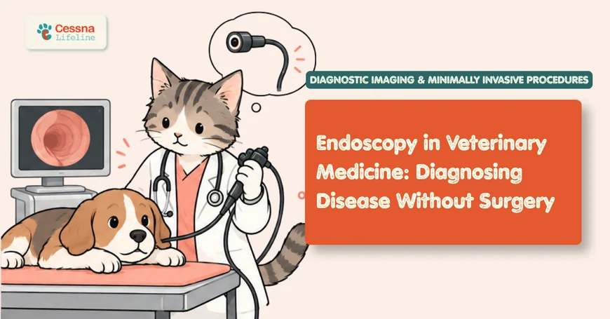
When a dog or cat has chronic vomiting, weight loss, or diarrhea that does not resolve with standard treatment, the next diagnostic step is often tissue: a biopsy that tells the pathologist whether the gut wall architecture is normal, inflamed, or infiltrated by neoplastic cells. For decades, getting that tissue required exploratory laparotomy, an open abdominal surgery with a week of restricted recovery. Endoscopy changed that calculus. A flexible endoscope passed through the mouth under general anesthesia can visualize the esophagus, stomach, and proximal duodenum, collect multiple targeted biopsies, retrieve foreign objects, and be completed in 30 to 60 minutes with a same-day discharge. vomiting in dogs causes and when to worry
What to Know Veterinary endoscopy provides direct visual assessment of mucosal surfaces and targeted biopsy without open surgery. Upper GI endoscopy is the primary diagnostic tool for chronic vomiting, weight loss, and suspected inflammatory bowel disease or GI lymphoma in dogs and cats. Foreign body retrieval succeeds in 85 to 95% of cases endoscopically. Recovery is measured in hours rather than weeks (Juvet et al., 2010).
The Scope That Sees What Surgery Cannot Find
An endoscope is a long, flexible tube containing a fiber-optic light source, a camera, and one or more working channels through which instruments can be passed: biopsy forceps, retrieval baskets, cytology brushes, foreign body graspers, and injection needles. The image from the camera tip is transmitted in real time to a monitor, allowing the endoscopist to navigate through the lumen while observing mucosa, recording video, and performing procedures simultaneously.
Veterinary endoscopy encompasses several distinct procedures depending on which body cavity or organ is examined:
Gastroscopy and upper GI endoscopy: The scope is passed per os (through the mouth) through the esophagus, into the stomach, and through the pylorus into the proximal duodenum. This is the most common veterinary endoscopic procedure and addresses the vast majority of upper GI diagnostic questions.
Colonoscopy: The scope is passed per rectum through the descending colon, transverse colon, ascending colon, cecum, and ileocolic junction. Used for large bowel disease: colitis, colorectal polyps, colorectal tumors, and chronic large-bowel diarrhea.
Rhinoscopy: A rigid or flexible scope is passed into the nasal passages to directly visualize the nasal mucosa, retrieve biopsy specimens from nasal masses, identify nasal foreign bodies (grass awns, plant material), and detect fungal plaques in aspergillosis. Often performed in conjunction with CT of the nasal cavity.
Bronchoscopy: The scope is passed through the larynx and trachea into the bronchi to visualize the lower airway, collect bronchoalveolar lavage (BAL) fluid for cytology and culture, and retrieve airway foreign bodies. Used in dogs and cats with chronic cough, hemoptysis, or suspected lower airway disease.
Cystoscopy: A rigid or flexible scope is passed through the urethra into the bladder to visualize the urothelium, collect biopsies from bladder masses, and assess the ureteral openings. Used for suspected transitional cell carcinoma (TCC), chronic urinary signs, and ectopic ureter assessment in female dogs.
The common thread across all these applications is that a camera reaches the target tissue through a natural orifice, eliminating the need for a surgical incision to access the diagnostic site.
What Conditions Can Endoscopy Diagnose in Dogs and Cats?
The diagnostic power of endoscopy lies in combining direct visual assessment of mucosal surfaces with targeted tissue sampling. Many GI conditions produce characteristic visual changes that an experienced endoscopist recognizes immediately; all require histopathological confirmation from biopsy specimens for definitive diagnosis. GI disease in dogs signs guide
Inflammatory bowel disease (IBD): The most common indication for upper GI endoscopy in dogs and cats with chronic vomiting and weight loss. IBD encompasses lymphoplasmacytic enteritis, eosinophilic enteritis, and other inflammatory infiltrates that are histologically distinguishable. The mucosa may appear grossly normal, granular, or erythematous with visible erosions; histopathology of biopsies provides the definitive diagnosis and the specific inflammatory subtype.
GI lymphoma: Low-grade alimentary lymphoma is the most common intestinal tumor in cats and is notoriously difficult to distinguish from IBD on visual inspection; the endoscopic mucosal appearance of the two conditions is often identical. Histopathology and immunohistochemistry (IHC) on endoscopic biopsies can distinguish them in many cases, though full-thickness surgical biopsies are sometimes required when superficial endoscopic samples are insufficient.
Gastric ulceration and erosion: NSAID-induced gastric ulcers, stress ulcers, and gastrinoma-associated hyperacid states produce characteristic gastric mucosal lesions visible on gastroscopy. Ulcer depth, location, and presence of active bleeding are assessed during the procedure.
Esophageal disease: Reflux esophagitis, esophageal strictures, esophageal foreign bodies, and vascular ring anomalies (in young dogs with regurgitation from birth) are all accessible to upper GI endoscopy. Strictures can be dilated during the same procedure using balloon dilators passed through the working channel.
Nasal disease: Rhinoscopy identifies nasal aspergillosis (fungal plaques on the turbinates), nasal foreign bodies, and nasal tumors through direct visualization and targeted biopsy of abnormal tissue.
Citation Capsule: Endoscopic biopsy of the duodenum and ileum (where accessible) in dogs with protein-losing enteropathy or chronic GI signs provides diagnostic histopathology in a high proportion of cases when a minimum of six to eight biopsies are collected from each anatomical site; sampling fewer sites or collecting fewer specimens significantly reduces diagnostic sensitivity for patchy diseases such as lymphoma and increases the risk of a false-negative result (Willard et al., 2008). The WSAVA GI Standardization Group guidelines mandate minimum biopsy numbers per site to ensure adequate diagnostic material from endoscopic procedures.
How Does a Veterinary Endoscopy Procedure Work?
Understanding the procedure sequence helps owners set accurate expectations for what the appointment involves and what the recovery looks like.
Preparation: Upper GI endoscopy requires complete gastric emptying; food is withheld for 12 to 18 hours prior to the procedure. Colonoscopy requires both dietary preparation (bland diet for 24 to 48 hours) and bowel preparation (enemas the evening before and morning of the procedure) to clear the colonic contents that would otherwise obscure the camera and contaminate biopsy specimens.
Anesthesia: General anesthesia is required for all endoscopic procedures in dogs and cats. An endotracheal tube protects the airway during upper GI endoscopy when the scope passes through the pharynx and esophagus. Positioning is typically left lateral recumbency for upper GI endoscopy.
Scope passage and navigation: The endoscope is passed through the mouth, into the esophagus, through the cardia (esophagogastric junction), into the stomach, and through the pylorus into the duodenum. Each anatomical transition requires specific technique: the pylorus in particular requires patience and appropriate scope positioning because it does not lie in a straight line from the gastric body.
Systematic mucosal assessment: The endoscopist examines each region systematically, documenting abnormalities in video and still images. The gastric rugae, antral mucosa, pyloric region, and duodenal villi each have characteristic normal appearances; abnormalities are described by location, extent, depth, and gross character.
Biopsy collection: Biopsy forceps are passed through the working channel and opened against the mucosa; a sample is collected with each closure. Multiple biopsies are collected from each anatomical region visited (duodenum, stomach body, antrum, esophagus as indicated). Samples are placed in formalin for histopathology.
Foreign body retrieval: If a foreign body is present in the esophagus or stomach and is amenable to retrieval, the appropriate instrument (basket, grasper, snare, or tripod forceps depending on object shape and material) is selected and the object is maneuvered out through the mouth under direct vision.
What veterinary gastroenterologists observe consistently: Owners frequently underestimate what endoscopic foreign body retrieval means relative to the surgical alternative when they first hear the recommendation. A dog that swallowed a rubber toy piece visible in the stomach on radiograph has approximately an 85 to 95% chance of retrieval under endoscopy without a single incision, under the same general anesthesia that any procedure requires. The alternative, an exploratory gastrotomy, involves abdominal incision, a 7 to 10 day course of antibiotics, a 2 to 3 week return-to-activity restriction, and a significantly higher risk of post-operative complications. When that comparison is made explicitly, the decision to proceed with endoscopy first is invariably straightforward for the owner. The recommendation to "try endoscopy before surgery" is not a hedge; it is the appropriate clinical sequence.
What Can Endoscopy Retrieve, Treat, and Sample?
Beyond biopsy collection, endoscopy provides several therapeutic capabilities that eliminate the need for surgical intervention:
Foreign body retrieval: Objects in the esophagus, stomach, and occasionally the proximal duodenum can often be retrieved under endoscopic visualization. The choice of retrieval instrument depends on the object: basket forceps for smooth rounded objects (balls, coins), tripod or alligator forceps for irregularly shaped objects (fabric, rubber), and polypectomy snares for linear foreign bodies such as string. Objects that are too large, too sharp to safely withdraw through the esophagus, or too deeply embedded in the mucosa may still require surgical extraction.
Esophageal stricture dilation: Strictures (narrowings) from previous esophageal foreign bodies, reflux, or surgery can be dilated using through-the-scope balloon dilators. The balloon is positioned across the stricture and inflated to a controlled pressure, stretching the fibrous scar tissue. Multiple sessions are often required for complete resolution.
Gastric polyp and colonic polyp removal: Pedunculated polyps can be snared and removed endoscopically (polypectomy) without surgical entry into the abdomen.
Nasal fungal treatment (aspergillosis): Rhinoscopy allows the endoscopist to visualize and mechanically debride fungal plaques from the nasal turbinates and flush antifungal medication into the nasal cavity under direct vision, which is superior to blind instillation. nasal tumor diagnosis dogs
Laparoscopy (keyhole surgery using small port incisions in the abdomen) is distinct from flexible GI endoscopy but falls under the broader endoscopy category in veterinary medicine. Its unique advantage over flexible GI endoscopy for GI diagnosis is that laparoscopic GI biopsies are full-thickness: the instrument passes through all layers of the bowel wall rather than only the mucosal surface. This is clinically important because some diseases, particularly lymphoma and pythiosis (a fungal GI infection in southern US dogs), primarily involve deeper bowel layers that superficial endoscopic biopsies do not reach. When ultrasound suggests marked bowel wall thickening or loss of wall layering in regions beyond endoscopic reach (jejunum, ileum), laparoscopy provides full-thickness biopsy of those areas through 3 to 5 mm port incisions rather than a full exploratory laparotomy incision.
Preparing Your Pet for Endoscopy: What to Expect
Endoscopy vs. Alternative Diagnostic Approaches: When to Choose Which
| Clinical Scenario | Best First Choice | Reason |
|---|---|---|
| Chronic vomiting + weight loss, normal radiographs | Upper GI endoscopy | Direct visualization + mucosal biopsy |
| Suspected gastric foreign body, accessible size | Endoscopic retrieval first | 85-95% success; avoids gastrotomy |
| Large bowel diarrhea + fresh blood | Colonoscopy | Mucosal assessment + biopsy of colon |
| Chronic nasal discharge, one side, CT performed | Rhinoscopy post-CT | Biopsy of mass identified on CT |
| Jejunal or ileal wall thickening on ultrasound | Laparoscopy or surgical biopsy | Beyond flexible endoscope reach |
| Suspected airway foreign body or chronic cough | Bronchoscopy | BAL + airway visualization |
| Marked intestinal wall thickening, loss of layering | Surgical/laparoscopic full-thickness biopsy | Superficial biopsy likely insufficient |
| Bladder mass suspicious for TCC | Cystoscopy + biopsy | Avoids open cystotomy |
The appointment: Owners should expect the procedure to run 1 to 3 hours from arrival to discharge, including pre-anesthetic preparation, the procedure itself, and recovery monitoring. Most animals are discharged the same day. Mild sedation-related drowsiness persists for 12 to 24 hours after discharge.
Post-procedure care: After upper GI endoscopy, food is withheld for 2 to 4 hours post-recovery before a small bland meal is offered. After colonoscopy, the colon requires 24 to 48 hours for the bowel preparation to fully clear. Animals with biopsy sites may show traces of blood in vomit or stool for 24 to 48 hours; persistent or heavy bleeding warrants prompt contact with the veterinary team. Activity restriction is minimal (leash walks for 24 hours) compared to the weeks required after open surgery.
Biopsy results: Histopathology results typically take 3 to 7 business days from tissue submission. Specific diagnoses requiring immunohistochemistry (IHC) or clonality testing (to distinguish IBD from lymphoma) may take 10 to 14 days. The referring veterinarian will contact the owner once results are available to discuss diagnosis and treatment plan.
How Accurate Is Endoscopy Compared to Other Diagnostics?
Endoscopy is highly accurate when the target lesion is within visual and biopsy reach and when adequate samples are collected. Its limitations are defined by reach and depth rather than by technique.
Within its reach, endoscopy is highly accurate: Gastric ulcers, esophageal lesions, and grossly abnormal mucosal disease are identified with high sensitivity on gastroscopy. Histopathological diagnosis from adequately collected endoscopic biopsies has high concordance with surgical biopsy for diseases that primarily involve the mucosal layer.
Outside its reach, it has defined limitations: The flexible endoscope passed orally reaches only to the proximal duodenum in most dogs (approximately 30 to 50 cm beyond the pylorus). The jejunum and ileum, which comprise the majority of the small intestinal length, are inaccessible to conventional upper GI endoscopy. Diseases that primarily affect these regions (pythiosis, jejunal lymphoma, some cases of protein-losing enteropathy) may return false-negative endoscopic biopsies. Ultrasound assessment of bowel wall architecture before endoscopy helps predict whether the lesion is likely to be within endoscopic reach. IBD in dogs and cats guide
Citation Capsule: A study comparing endoscopic mucosal biopsies to full-thickness surgical biopsies in the same dogs with chronic GI signs found that the two methods agreed on diagnosis in approximately 70 to 80% of cases; disagreement occurred predominantly in cases where the primary pathology involved the submucosa or muscularis propria layers inaccessible to mucosal biopsy forceps, particularly in dogs with small cell lymphoma and pythiosis (Kleinschmidt et al., 2006). This concordance rate supports endoscopic biopsy as the appropriate first-line diagnostic approach for most cases, with full-thickness surgical biopsy reserved for endoscopy-negative cases with persistent clinical signs.
An emerging advance in veterinary endoscopy is chromoendoscopy and narrow band imaging (NBI). Applied at the time of endoscopy, chromoendoscopy involves spraying dilute dye (methylene blue or indigo carmine) onto the mucosal surface to highlight subtle texture changes; narrow band imaging uses specific light wavelengths that enhance mucosal vascular patterns. Both techniques, standard in human GI oncology for flat polyp detection and IBD severity grading, are beginning to be used in veterinary medicine to detect subtle mucosal changes that appear grossly normal under standard white light. They allow the endoscopist to target biopsies more precisely to abnormal areas rather than distributing samples randomly across a grossly normal mucosa, potentially improving diagnostic yield for early or patchy disease.
Citation Capsule: Endoscopic biopsy of the proximal small intestine and stomach in dogs with chronic vomiting and weight loss achieves diagnostic histopathology in a high proportion of cases when the standardized WSAVA biopsy protocol is followed; the most common cause of non-diagnostic endoscopic biopsy is collection of an insufficient number of specimens, use of inappropriately sized biopsy forceps, or failure to include duodenal biopsies in a protocol limited to the stomach (Willard et al., 2008). Protocol adherence is the single most important controllable variable in endoscopic diagnostic yield.
Does Endoscopy Work Differently in Cats?
The fundamental technique is the same in cats as in dogs, but feline anatomy and the specific diseases that cause GI signs in cats create meaningful differences in practice.
Scope size: Cat GI anatomy is substantially smaller than dogs. Smaller diameter scopes (pediatric or small animal-specific instruments) are required for feline upper GI endoscopy. Standard adult dog endoscopes cannot negotiate the feline pylorus or duodenum safely.
IBD versus lymphoma in cats: The most clinically significant challenge in feline GI endoscopy is distinguishing low-grade alimentary lymphoma (LGAL) from IBD. Both conditions are common in middle-aged to older cats, both cause chronic vomiting and weight loss, and both can present with grossly normal endoscopic mucosa. The distinction requires histopathology and immunohistochemistry (IHC) to assess lymphocyte immunophenotype, and in some cases clonality testing. The two conditions have different treatments and different prognoses: IBD is managed with diet and immunosuppression; LGAL is managed with chlorambucil and prednisolone chemotherapy with median survival times exceeding 2 years with appropriate treatment.
Cats and colonoscopy: Colonoscopy in cats requires the same bowel preparation as dogs, but the smaller colonic diameter and the cat's tendency to be less cooperative during recovery require experienced handling. Feline large bowel disease (colitis, colorectal polyps) is less common than in dogs but represents an important subset of cats with chronic large-bowel diarrhea who benefit from colonoscopic assessment. lower GI disease in dogs colonoscopy
Frequently Asked Questions
Does pet endoscopy require general anesthesia?
Yes. Unlike human endoscopy, where patients remain awake or lightly sedated, dogs and cats cannot cooperate with scope passage through the pharynx and esophagus. General anesthesia ensures patient safety during scope navigation, prevents reflex gagging and airway contamination, and allows the endoscopist to work methodically without the stress of patient movement. The endotracheal tube placed during anesthesia for upper GI endoscopy also protects the airway if any gastric contents are encountered.
How long does a pet endoscopy take?
The procedure itself takes 20 to 60 minutes depending on scope type and whether biopsies or foreign body retrieval is performed. Total appointment time, including pre-anesthetic preparation, anesthetic induction, the procedure, and recovery, is typically 2 to 4 hours. Most pets are discharged the same day.
What is the difference between endoscopy and exploratory surgery for GI diagnosis?
Endoscopy provides mucosal-level biopsy through a natural orifice without incisions; recovery is hours and complications are uncommon. Exploratory surgery (laparotomy) provides access to the full abdominal cavity, full-thickness GI biopsy, and direct visualization of all organs; recovery is weeks and carries higher risk. Endoscopy is the appropriate first-line diagnostic approach when the suspected disease involves the mucosal layer of accessible regions. Surgery is required when the lesion is beyond endoscopic reach, when full-thickness tissue is needed, or when concurrent surgical treatment (tumor removal, intestinal resection) is planned.
What happens if the foreign body cannot be retrieved endoscopically?
If an object cannot be safely retrieved through the endoscope (too large, too sharp, embedded in the mucosa, or in a position that makes withdrawal hazardous), the endoscopist terminates the attempt without having harmed the patient or worsened the situation. Surgical retrieval is then performed. The endoscopic attempt does not compromise the surgical option. In some cases, the endoscopist can advance the object past the pylorus into the small intestine if it cannot be retrieved orally, allowing it to pass naturally rather than requiring gastrotomy.
What conditions does endoscopy find that other tests miss?
Endoscopy provides the only way to directly visualize mucosal surface changes (erosions, ulcers, granularity, nodularity, vascular pattern changes) and to collect targeted tissue samples from specific anatomical sites. Blood work, imaging, and ultrasound can identify organ involvement, wall thickening, or mass lesions, but none provides tissue diagnosis at the mucosal level. IBD versus GI lymphoma, for example, cannot be distinguished without histopathology from tissue samples; endoscopy collects those samples with the least patient morbidity of any technique that reaches the target site. foreign body ingestion dogs emergency
The Answer That Was Already Inside
Endoscopy is, at its core, a way to ask the body directly what is wrong. Not from outside, not through the walls, not inferred from blood values or ultrasound shadows, but by sending a camera to the site of the problem and looking. The tissue biopsy it returns answers the question that clinical signs raise but cannot answer on their own: is this inflammation, is this lymphoma, is this something that responds to steroids or to chemotherapy or to diet alone.
For the dog or cat with months of weight loss and vomiting and a stack of inconclusive test results, endoscopy frequently provides in a single 40-minute procedure under anesthesia what all the prior testing approached but could not reach. That efficiency is not incidental to endoscopy's value. It is most of the value.