CT Scans for Pets: When Advanced Imaging Is Needed
A veterinary CT scan shows what X-rays cannot. Learn when dogs and cats need CT imaging, what the procedure involves, and which conditions it diagnoses best.
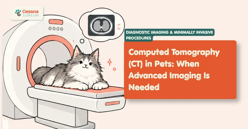
Plain radiographs have been the backbone of veterinary imaging for decades, and they remain indispensable. But there are clinical questions radiographs cannot answer, anatomical territories they cannot resolve, and treatment decisions that cannot be made with confidence from a two-dimensional image of a three-dimensional structure. Computed tomography fills that diagnostic gap. In veterinary medicine, CT has moved from a specialty novelty to an accessible and increasingly expected standard of care for complex diagnostic and surgical planning scenarios. veterinary specialist referral guide
What to Know A veterinary CT scan acquires hundreds of cross-sectional images in minutes, reconstructed into three-dimensional anatomical maps with sub-millimeter resolution. General anesthesia is required because even minor patient movement renders the images uninterpretable. CT is the preferred modality for bone, nasal cavity, thoracic, and many orthopedic assessments. MRI remains superior for brain parenchyma and spinal cord soft tissue. Cost typically ranges from $1,200 to $3,000 including anesthesia.
The Imaging Tool That Changed Veterinary Diagnosis
Computed tomography works on the same physical principle as plain radiography: X-rays are attenuated differently by tissues of different densities. What CT adds is acquisition geometry. Instead of a single X-ray beam passing through the patient to a flat detector plate, a CT scanner rotates an X-ray source continuously around the patient while an array of detectors on the opposite side records attenuation at hundreds of angles. A computer reconstructs these attenuation measurements using filtered back-projection algorithms to generate a series of cross-sectional "slices" through the patient's body.
Modern multi-detector CT scanners (MDCT) can complete a full helical scan of the thorax in under 10 seconds, capturing images during a single breath-hold equivalent and dramatically reducing motion artifact. A full body scan that took 45 minutes on first-generation CT equipment now takes 3 to 8 minutes of actual scan time. The reconstructed images can be manipulated post-acquisition to view the anatomy in any plane (axial, sagittal, coronal) or rendered as a three-dimensional volume, and specific tissue windows can be applied to emphasize bone detail, soft tissue contrast, or vascular structures depending on the diagnostic question.
The key quantitative output of CT is the Hounsfield unit (HU), a standardized scale of tissue radiodensity. Air registers at approximately minus 1,000 HU; water at 0 HU; soft tissue at 20 to 80 HU; bone at 400 to 1,000 HU. This numerical scale allows the radiologist to characterize tissue composition with a specificity not achievable from the qualitative interpretation of a grey-scale radiograph. A soft tissue mass measuring 60 HU behaves differently from one measuring 10 HU, and that difference is diagnostically meaningful for distinguishing abscess from neoplasia, for example.
Contrast agents (iodinated contrast medium, administered intravenously) enhance vascular structures and alter the HU of tissues that have disrupted or hyperpermeable blood supply, such as tumors, abscesses, and infarcts. Contrast-enhanced CT is used for the majority of oncological, vascular, and soft tissue diagnostic CT studies.
How Does a CT Scanner Work and What Does It Show?
The tissues CT visualizes best are those with the highest inherent contrast: bone, air-containing structures, and the interfaces between them. This is why CT excels in specific anatomical regions and for specific diagnostic questions.
Bone: CT resolves bony architecture at sub-millimeter resolution, allowing detection of subtle cortical lysis, endosteal new bone formation, joint surface changes, and fracture fragment orientation that is partially or completely obscured on radiographs by overlying structures. Skull base, nasal cavity, tympanic bullae (ear canal), vertebral bodies, elbow and shoulder joints, and the carpus and tarsus are all anatomical areas where CT provides substantially superior bone detail compared to radiographs.
Air-filled structures: The nasal cavity, paranasal sinuses, trachea, bronchi, and pulmonary parenchyma are naturally high-contrast on CT because air (minus 1,000 HU) provides extreme density difference from adjacent soft tissue (20 to 80 HU). This is why pulmonary CT is so sensitive for small nodules, subtle bronchial changes, and pleural abnormalities that are invisible on thoracic radiographs.
Soft tissue: CT distinguishes soft tissue structures less well than MRI, but contrast enhancement significantly improves soft tissue characterization for vascular tumors, lymph nodes, organ masses, and peritoneal detail. The liver, spleen, adrenal glands, and kidneys are assessable on abdominal CT, and contrast enhancement patterns help distinguish benign from malignant lesions.
Vascular structures: CT angiography (CTA), using rapid post-contrast acquisition during the arterial phase, produces detailed images of major vessels and their branches. This is the technique used to detect pulmonary thromboembolism, portosystemic shunts, and vascular malformations in dogs and cats. pulmonary thromboembolism dogs
The nasal cavity and paranasal sinuses represent the anatomical territory where CT is most dramatically superior to all alternative imaging modalities. Three factors converge: the nasal cavity contains air (maximally CT-bright), bone (maximally CT-white), and soft tissue (intermediate CT-grey), producing inherently high-contrast images where lesion boundaries are precisely delineated. Radiographs of the same region produce summation shadows from overlapping nasal turbinates, incisive bones, and maxillary structures that render the margin of even a large nasal tumor ambiguous. CT converts a radiographic description of "increased nasal opacity with possible turbinate destruction" into a surgical map showing the exact extent of tumor, which walls are involved, whether the cribiform plate is intact (critical for radiation planning), and whether the tumor has crossed the midline. This is why CT is considered the minimum imaging standard before any nasal tumor treatment decision.
Citation Capsule: Multi-detector CT scanners complete helical thoracic acquisition in less than 10 seconds in dogs and cats, capturing images that are virtually free of respiratory motion artifact and providing cross-sectional pulmonary anatomy at resolutions of 0.5 to 1.0 mm slice thickness, enabling detection of pulmonary nodules as small as 3 to 5 mm that are below the detection threshold of thoracic radiographs (Reetz et al., 2006). This sensitivity difference is clinically significant for oncological staging, where small pulmonary metastases detected on CT but not radiographs alter the treatment plan and prognosis in a meaningful proportion of cases.
When Does a Dog or Cat Actually Need a CT Scan?
The clinical scenarios where CT provides diagnostic information that changes the treatment plan fall into several distinct categories. The question is not "would CT be interesting" but "would the result change what we do."
Nasal cavity and skull: Any dog or cat with chronic unilateral epistaxis (nosebleed), persistent unilateral nasal discharge unresponsive to antibiotics, facial deformity, or exophthalmos (eye protrusion from a retrobulbar mass) should be considered for CT before biopsy or treatment. CT defines tumor extent, identifies cribiform plate integrity, and determines radiation field parameters. nasal tumor in dogs
Thorax and pulmonary staging: CT pulmonary staging is significantly more sensitive than thoracic radiographs for detecting small pulmonary metastases. Many oncologists now recommend thoracic CT rather than radiographs for staging of primary tumors with pulmonary metastatic potential (osteosarcoma, mast cell tumors, soft tissue sarcomas, mammary carcinoma). CT pulmonary angiography (CTPA) is also the gold standard for diagnosing pulmonary thromboembolism in dogs.
Brain and spinal column: CT provides excellent visualization of skull and vertebral bone, making it the preferred first-line study for fractures, disc material mineralization, and mass lesions with bony involvement. However, MRI surpasses CT for brain parenchyma, spinal cord, and nerve root detail (discussed in the next section).
Orthopedic joints: Elbow dysplasia, shoulder OCD, complex intra-articular fractures, and planning for triple pelvic osteotomy (TPO) all benefit from CT. The joint is imaged in all planes simultaneously, and three-dimensional reconstructions allow the surgeon to visualize fragment size, position, and displacement in a way that multi-view radiographs cannot match. orthopedic surgery planning dogs
Ear canal and tympanic bulla: Otitis media and interna, cholesteatoma, and nasopharyngeal polyps involving the middle ear in cats are assessed by CT of the tympanic bullae with far greater sensitivity than skull radiographs. The bullar wall thickness, fluid content, ossicular chain integrity, and extent of any soft tissue mass are all assessable.
Abdominal surgical planning: CT of the abdomen is used for adrenal tumor assessment (invasion of caudal vena cava is a critical pre-surgical finding), portosystemic shunt mapping (to determine shunt anatomy before surgical attenuation), and large mass characterization when ultrasound is indeterminate.
Citation Capsule: Thoracic CT detects pulmonary nodules in dogs with known primary tumors at a significantly higher rate than thoracic radiographs: in one study, CT identified pulmonary lesions in dogs staged as radiographically node-negative, upstaging a clinically meaningful proportion of cases and altering surgical or chemotherapy planning (Reetz et al., 2006). This sensitivity gap has driven many veterinary oncologists to recommend thoracic CT as the preferred staging modality for primary tumors with pulmonary metastatic potential.
What Happens During a Veterinary CT Scan?
Understanding the procedure helps owners prepare and set accurate expectations. A veterinary CT scan appointment typically proceeds through the following phases:
Pre-anesthetic assessment: The animal is examined, blood work is reviewed (or collected on the day for elective procedures), and body weight is recorded for anesthetic and contrast dosing. Intravenous catheter placement is standard. An empty stomach is required, typically 8 to 12 hours of fasting before general anesthesia.
Anesthetic induction: General anesthesia is induced and maintained throughout the scan. An endotracheal tube is placed and the patient is connected to a circuit delivering inhalant anesthetic and oxygen. Respiratory rate and depth may be controlled by the anesthetist to time image acquisition during breath-holding phases for thoracic CT, reducing respiratory motion artifact.
Patient positioning and scan acquisition: The patient is placed on the CT table in sternal or dorsal recumbency depending on the region being scanned. Scout images (topograms) are acquired first, and the scan field is selected. The table advances through the scanner gantry while the X-ray source rotates. Modern MDCT completes the diagnostic scan in 2 to 15 minutes depending on body region and slice thickness. The patient does not move; the CT table and rotating gantry do all the work.
The reason veterinary CT requires general anesthesia, when human CT does not, is not institutional conservatism or a lower tolerance for inconvenience. It is image physics. The sub-millimeter slice thicknesses required for diagnostic CT of the skull and nasal cavity (typically 0.5 to 1.0 mm) mean that a movement of one millimeter during acquisition renders the reconstructed image uninterpretable: the small lesion that needs to be distinguished from normal anatomy cannot be separated from motion artifact. A dog or cat breathing quietly under light sedation moves its skull approximately 2 to 5 mm with each respiratory cycle. A dog lying still under sedation moves periodically during swallowing, startling, or normal fluctuations in sedation depth. General anesthesia with a secured airway allows the anesthetist to control respiratory rate and depth, stop breathing on command during critical sequences, and maintain consistent positioning for the entire scan. A technically perfect 8-minute scan under general anesthesia is diagnostically superior to a 2-minute scan of a sedated but breathing dog.
Contrast administration (if indicated): Iodinated contrast medium is administered intravenously by the anesthetic team at a specific dose and timing relative to the scan acquisition, depending on whether arterial, portal venous, or delayed-phase imaging is required. Post-contrast sequences are acquired immediately following contrast injection.
Recovery: The patient is extubated once swallowing reflexes return, moved to a recovery area, and monitored until sternal and ambulatory. Total time from anesthetic induction to recovery ward is typically 30 to 60 minutes. Most patients are discharged the same day unless concurrent procedures (such as a CT-guided biopsy or concurrent surgery) are performed.
Image review and reporting: Images are reviewed by a veterinary radiologist (specialist in veterinary diagnostic imaging), who provides a written report to the referring veterinarian. Reports typically include systematic description of all findings, interpretation, differential diagnoses, and imaging recommendations.
CT Versus MRI: Choosing the Right Modality
CT and MRI are complementary modalities, not competitors, but they have distinct tissue sensitivities that determine which is appropriate for a given clinical question.
Imaging Modality Comparison: CT, MRI, Radiograph, Ultrasound
| Feature | CT | MRI | Radiograph | Ultrasound |
|---|---|---|---|---|
| Best tissue type | Bone, air, vessels | Soft tissue, brain, spinal cord | Bone, lungs, gross soft tissue | Abdominal organs, cardiac, guided biopsy |
| Spatial resolution | Sub-mm (0.5 mm) | 0.5-1.5 mm | 0.1-0.3 mm | 0.5-2 mm |
| Ionizing radiation | Yes | No | Yes (lower dose) | No |
| Anesthesia required (pets) | Yes | Yes | Usually not | Usually not |
| Typical scan duration | 2-15 min | 30-90 min | Minutes | 15-45 min |
| Approximate cost (US) | $1,200-$3,000 | $1,800-$4,000 | $150-$400 | $300-$700 |
| Primary veterinary strength | Nasal, bone, thorax, vascular | Brain, spinal cord, joints | Lungs, fractures, initial screen | Organ masses, cardiac, FAST |
CT is preferred over MRI for:
- Nasal cavity and paranasal sinus disease
- Skull and tympanic bulla assessment
- Thoracic CT and pulmonary staging
- Bony vertebral column (fractures, disc mineralization, osteolysis)
- Vascular assessment (CT angiography)
- Orthopedic surgical planning (joints, fractures)
- Abdominal organ surgical planning where MRI is unavailable
MRI is preferred over CT for:
- Brain parenchyma (gray/white matter, intracranial lesions, hydrocephalus)
- Spinal cord and nerve root detail (intramedullary vs. extramedullary lesion differentiation)
- Pituitary gland (pituitary-dependent hyperadrenocorticism)
- Soft tissue tumors requiring precise margin mapping for surgery
- Perineural invasion assessment
Citation Capsule: MRI provides substantially superior soft tissue contrast compared to CT for brain parenchyma assessment; the signal-to-noise ratio for gray-white matter differentiation and the ability to detect subtle changes in water content (T2 hyperintensity) within brain tissue make MRI the preferred modality for intra-axial lesions, inflammatory encephalitis, and cortical infarcts in dogs and cats (Garosi, 2010). CT of the brain is reserved for acute trauma (to detect hemorrhage rapidly), bony calvarium lesions, and situations where MRI is unavailable or the scan duration is clinically prohibitive.
What Do CT Results Look Like and How Are They Used?
CT results are delivered to the referring veterinarian as a written radiologist report accompanied by a CD or digital transfer of the DICOM image files. The report describes findings systematically by body region, provides a differential diagnosis list ranked by probability, and makes imaging recommendations where relevant.
What veterinary radiologists observe consistently: Approximately 30 to 40% of thoracic CT scans performed for oncological staging in dogs reveal findings that were not detected on same-day thoracic radiographs. These include small pulmonary nodules (below the 5 to 8 mm radiographic detection threshold), tracheobronchial lymphadenopathy, pleural thickening, and mediastinal masses. In a meaningful proportion of these cases, the CT finding changes the oncological stage and, with it, the treatment recommendation: a dog planned for curative-intent surgery based on radiographically clean lungs may instead receive a palliative protocol when CT reveals multiple sub-centimeter pulmonary nodules. The clinical implication is that for cancers with high pulmonary metastatic potential, "clear chest radiographs" and "clear chest CT" are not equivalent statements. dog cancer diagnostic workup
Understanding CT reports requires recognizing their vocabulary. Findings described as "hyperdense" on CT are brighter than surrounding tissue on the greyscale display (more X-ray attenuating: bone, contrast-enhanced vessels, acute hemorrhage). "Hypodense" findings are darker (less attenuating: fat, necrosis, fluid, air). "Heterogeneous enhancement" after contrast means the lesion has irregular internal vascularity, which is associated with malignancy and necrosis. "Lytic" bone lesions have lost cortical density; "productive" lesions have added new bone.
Citation Capsule: CT-guided fine needle aspiration and core biopsy of thoracic, abdominal, and skull-base masses allows real-time imaging confirmation of needle placement within the target tissue, significantly improving diagnostic yield compared to ultrasound-guided or blind biopsy of lesions that are not directly accessible or visible under ultrasound (Samii et al., 2004). CT-guided biopsy is particularly valuable for nasal masses, pulmonary nodules, and deep lymph nodes where ultrasound visualization is limited by bone, air, or depth.
Are There Risks, and How Much Does a Pet CT Scan Cost?
Radiation exposure: CT delivers significantly more radiation than plain radiographs. A single thoracic CT in a dog delivers approximately 1 to 5 mGy (milligray) of radiation dose, compared to less than 0.1 mGy for a two-view thoracic radiograph. For a single diagnostic CT event, the radiation dose is not considered clinically significant for the patient. In animals with chronic disease requiring repeated CT studies over months, cumulative exposure should be considered when selecting intervals and whether alternative modalities are appropriate.
Contrast reactions: Iodinated contrast medium carries a small risk of adverse reaction in any patient, ranging from mild (nausea, vomiting) to severe (anaphylaxis). Contrast nephropathy (kidney injury) is a concern in patients with pre-existing renal insufficiency; hydration and renal function should be evaluated before contrast administration in at-risk patients. Overall adverse reaction rates to modern non-ionic iodinated contrast in veterinary patients are low, estimated at less than 1% for minor reactions and substantially lower for severe reactions.
Anesthetic risk: Because veterinary CT requires general anesthesia, the risk profile of the procedure is largely that of the anesthetic event rather than the CT scan itself. Animals with significant cardiac, respiratory, or metabolic disease require careful pre-anesthetic assessment, and the risk-benefit decision for the CT study is primarily an anesthetic risk calculation in these patients.
Cost: CT scans in veterinary medicine typically cost between $1,200 and $3,000 in the United States, including anesthetic and monitoring. The fee varies by institution (university teaching hospital versus private specialty center), geographic region, body region scanned, whether contrast is included, and whether the study is interpreted by an on-site radiologist or submitted for remote teleradiology reporting. Some pet insurance policies cover advanced imaging under the diagnostics or referral benefit; coverage confirmation before booking is recommended.
Citation Capsule: Iodinated contrast medium used in veterinary CT is a non-ionic, low-osmolarity formulation with a substantially improved safety profile compared to earlier ionic agents; adverse reaction rates in veterinary patients are estimated below 1% for minor reactions (vomiting, salivation) and substantially lower for severe anaphylactoid reactions, though pre-existing renal insufficiency remains a contraindication requiring individualized risk-benefit assessment before contrast administration (Pollard et al., 2008). Adequate pre-procedure hydration reduces nephrotoxic risk in patients with borderline renal function.
Frequently Asked Questions
Does a pet CT scan require anesthesia?
Yes. Unlike human CT, where patients can hold still on command, dogs and cats move unpredictably during scanning. Even a small amount of movement during the high-resolution sequences required for brain and nasal CT produces motion artifact that renders the images uninterpretable. General anesthesia ensures the patient is completely still, allows respiratory control during thoracic acquisition, and maintains consistent positioning throughout the study. The anesthesia required for CT carries its own risk, which is evaluated during the pre-scan assessment.
How long does a pet CT scan take?
The actual scanning time is short: 2 to 15 minutes depending on body region and slice thickness. However, the total appointment time is longer due to pre-anesthetic preparation, catheter placement, anesthetic induction, patient positioning, recovery monitoring, and discharge. Most CT appointments run 1 to 3 hours from arrival to discharge, depending on whether contrast is used, whether a CT-guided biopsy is performed concurrently, and how quickly the patient recovers from anesthesia.
What is the difference between a CT scan and an X-ray for a pet?
Both use X-rays, but a plain radiograph captures a single two-dimensional projection through the patient, where all internal structures are superimposed on one plane. CT acquires hundreds of projections at different angles around the patient and reconstructs them into cross-sectional slices, producing three-dimensional anatomical data at sub-millimeter resolution. CT eliminates the superimposition problem, distinguishes tissues by precise Hounsfield unit values rather than qualitative grey-scale, and allows post-acquisition reconstruction in any imaging plane or as a 3D volume.
When should I ask my vet about a CT scan for my dog?
Ask when your dog has a problem that plain radiographs or ultrasound have not explained, or when you have been told that the answers needed for treatment planning require advanced imaging. Specific triggers include: chronic unilateral nosebleed or nasal discharge, suspected nasal tumor, a mass in the chest that needs surgical planning, a brain or spinal cord problem suspected after neurological examination, complex joint disease in the elbow or shoulder, ear disease suspected to involve the middle ear, and cancer staging where pulmonary metastases need to be definitively excluded. Your veterinarian or a veterinary specialist will discuss whether the diagnostic gain from CT justifies the anesthetic procedure for your specific pet.
Can a CT scan detect cancer in dogs?
CT can detect masses, lymph node enlargement, pulmonary nodules, bone lysis, and vascular abnormalities consistent with cancer, and it characterizes their extent and relationship to surrounding structures in detail that changes treatment planning. However, a CT scan cannot confirm that a lesion is malignant: that determination requires tissue sampling (biopsy or fine needle aspiration) with histopathology or cytology. CT is a structural imaging tool; it shows anatomy and pathological changes but does not diagnose the biological nature of those changes without pathological confirmation. CT-guided biopsy can combine imaging precision with tissue sampling in a single procedure.
The Scan That Answers What the Exam Cannot
There are clinical questions that a thorough physical examination, well-positioned radiographs, and skilled ultrasound simply cannot answer: whether the cribiform plate is eroded, whether the pulmonary nodule seen radiographically is one or nine, whether the shunt is intrahepatic or extrahepatic, whether the adrenal mass has invaded the caudal vena cava. These are not questions about diagnostic acuity; they are questions about the physical limits of what different imaging modalities can resolve.
CT does not replace clinical judgment. It informs it, with a specificity and anatomical detail that allows treatment decisions to be made from a position of genuine knowledge rather than diagnostic inference. That is the reason it has become, in the right clinical context, not an optional advanced study but the minimum standard for making the treatment plan correctly. MRI for pets