Foreign Body in Dogs: Symptoms, Obstruction, and Treatment
Up to 20% mortality when intestinal perforation occurs from a swallowed foreign body. Learn dog swallowed object symptoms, obstruction signs, and treatment.
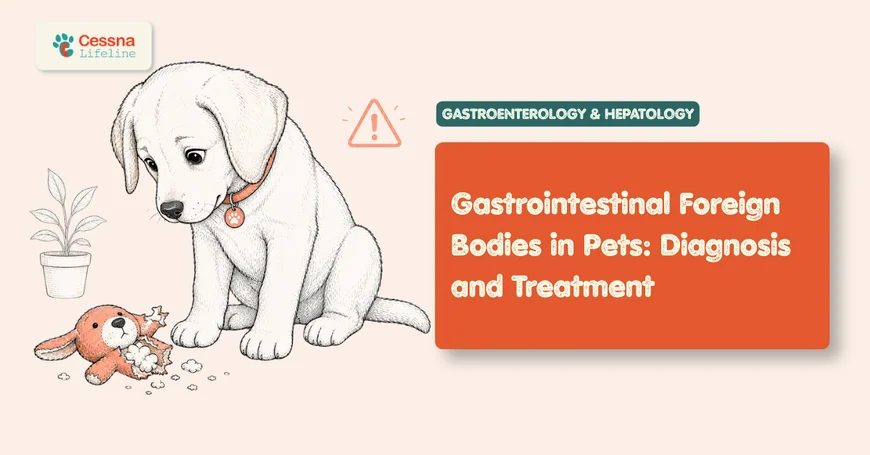
When a dog swallows something it shouldn't, the outcome depends almost entirely on what was ingested and how quickly it becomes a problem. Most objects that reach the stomach pass without incident. But for the fraction that become lodged in the intestine, complications escalate within hours. When intestinal perforation occurs, mortality can reach 20 to 70% even with surgical intervention (Ellison GW, Veterinary Surgery, 2011). Understanding dog swallowed object symptoms before that threshold is crossed is the difference between a straightforward endoscopic retrieval and emergency abdominal surgery with a guarded prognosis.
What to Know A foreign body in dogs becomes life-threatening when it causes intestinal obstruction or perforation. Most symptomatic dogs present with vomiting within hours of ingestion. Diagnosis relies on radiographs combined with abdominal ultrasound. Treatment ranges from monitored passing, to endoscopic removal, to surgical intervention depending on object location, type, and degree of obstruction. Linear objects (string, rope toy fibers, pantyhose) carry the highest risk of intestinal perforation and should never be managed with a "watch and wait" approach regardless of how mild early symptoms appear.
gastrointestinal emergencies in pets
How Does a Swallowed Object Become an Emergency?
Swallowing a foreign object is not itself the crisis. The crisis is what happens next, and that depends almost entirely on whether the object moves.
Dogs are indiscriminate eaters, and their anatomy tolerates a surprising range of swallowed items. A dog's esophagus, stomach, and intestinal tract are designed for processing whole prey items and rough plant matter. Objects that are small relative to the intestinal lumen, smooth, and non-toxic often traverse the entire GI tract without incident, appearing in stool within 24 to 72 hours.
The problem begins when an object is too large to exit the stomach, too rigid to conform to the intestinal curves, or too fibrous to compress. Gastric outflow obstruction occurs at the pylorus, the narrow junction between the stomach and the small intestine. Intestinal obstruction most frequently occurs in the jejunum, the longest and most mobile segment of the small intestine, where the lumen narrows and natural curves create lodging points.
The cascade once an obstruction develops is well understood. Fluid and gas accumulate proximal to the blockage. The distended bowel wall loses perfusion. Mucosal cells die. Bacteria translocate across the compromised wall. What started as an object stuck in the intestine becomes septic peritonitis. In retrospective studies of canine intestinal obstruction, the window between initial symptom onset and irreversible intestinal compromise is often measured in hours to one to two days (Merck Veterinary Manual, Gastrointestinal Obstruction in Small Animals, 2024). The later a dog reaches veterinary care in that window, the fewer surgical options remain.
when to take your dog to the emergency vet
What Are the Symptoms of a Foreign Body in Dogs?
Vomiting is the defining symptom of a foreign body in dogs, occurring in approximately 80% of cases with confirmed obstruction (Hayes G, Journal of Small Animal Practice, 2009). In partial obstruction, vomiting may be intermittent while some material still passes; in complete obstruction, it becomes repetitive and bilious. Dogs often attempt to eat and drink between vomiting episodes, which typically worsens the frequency.
The timing of vomiting relative to ingestion matters. Vomiting that begins immediately or within one to two hours of ingestion may indicate esophageal lodging, which requires separate assessment from intestinal obstruction. Vomiting that begins 6 to 12 hours after ingestion and escalates in frequency and severity is more consistent with pyloric or small intestinal obstruction.
Other symptoms of dog swallowed object that owners observe include:
- Abdominal discomfort ranging from mild guarding to severe pain on palpation
- Reduced or absent appetite following the initial vomiting onset
- Progressive lethargy as the obstruction advances
- Bloated or visibly distended abdomen, more prominent with complete obstruction
- Absence of defecation or straining without result
- Drooling, repeated lip-licking, or exaggerated swallowing if the object is in the esophagus
One symptom that owners frequently miss is what veterinarians describe as positional discomfort: the dog that keeps shifting, is reluctant to lie flat, or repeatedly stretches into a bowing position. Unlike the characteristic prayer position associated with pancreatitis, the dog with a foreign body tends to move restlessly without settling. The abdomen may feel firm or tense even before dramatic distension is visible.
The rate of deterioration varies with object type and location. A dog with a smooth rubber toy lodged in the jejunum can maintain relatively normal behavior for 12 to 18 hours before rapid decline. A dog with a corn cob or linear foreign body may deteriorate within 4 to 8 hours of lodging.
Where Foreign Bodies Get Stuck: Location Determines Severity
Not all lodging sites carry equal risk. The esophagus, stomach, small intestine, and large intestine present distinct clinical pictures, different diagnostic challenges, and very different treatment approaches. In a retrospective study of 208 canine GI foreign body cases, the jejunum was the most common intestinal lodging site, with socks ranking as the most frequently reported ingested object overall (Hayes G, Journal of Small Animal Practice, 2009).
Esophageal foreign bodies typically cause obvious distress immediately: pawing at the mouth, gagging, retching, drooling, and refusal to eat or drink. Bones, sticks, and large chunks of rawhide are common culprits. Esophageal perforation carries high morbidity, and most esophageal foreign bodies require endoscopic or surgical removal within hours of diagnosis.
Gastric foreign bodies represent the most favorable scenario for intervention. The stomach is a large, accessible reservoir where objects can sit for extended periods without immediately causing vascular compromise. Endoscopic retrieval is frequently possible for objects confirmed to be within the stomach, particularly if attempted within 24 to 48 hours of ingestion. Smooth, blunt objects in the stomach of a non-symptomatic dog may sometimes be managed with monitored observation plus serial radiographs.
Small intestinal obstruction carries the highest risk of the major locations. The small intestine has limited tolerance for distension-related blood supply compromise, and mucosal damage can occur within hours of complete obstruction. Jejunal obstructions are most common due to the length and curvature of this segment. Duodenal obstruction, just past the pylorus, is less common but particularly serious due to proximity to the bile and pancreatic ducts.
Colonic foreign bodies are less common, as most objects that reach the large intestine have already navigated the narrow points of the small intestine. Objects that do lodge in the colon often cause straining, tenesmus, and abnormal stool appearance, and may sometimes be managed with manual extraction if positioned favorably.
Corn cobs deserve particular attention because their texture is uniquely deceptive on radiographs. Unlike a ball or rigid toy, a corn cob has a diameter that roughly approximates the canine intestinal lumen, causing it to conform partially to the intestinal shape rather than presenting as a discrete lodged mass. The classic "sentinel loop" gas accumulation pattern on radiograph may not develop as dramatically as with other foreign bodies. This means corn cob obstruction diagnoses are occasionally delayed until the dog is significantly more compromised than initial imaging suggested, which is why clinical suspicion in a symptomatic dog with corn cob access should remain high even with inconclusive imaging.
<figcaption>Surgical mortality risk for canine foreign body removal increases significantly when intestinal perforation or peritonitis are present at the time of surgery. Early intervention keeps outcomes in the lower two categories.</figcaption> </figure>
How Do Vets Diagnose a GI Obstruction?
The diagnostic workup for a suspected foreign body in dogs begins with physical examination and moves rapidly to imaging. Abdominal palpation may reveal pain localization, a palpable mass, or tense distension. However, diagnosis cannot rest on physical examination alone, since many lodged objects are not palpable and pain severity varies considerably between individual dogs.
Abdominal radiographs are the first imaging step. Radio-opaque objects (metal, dense bone, some rubber toys) are directly visible. Radio-lucent objects (fabric, corn cobs, soft plastic, most rubber) require identification through secondary findings: asymmetric gas accumulation, distended intestinal loops, and loss of normal serosal detail. Radiograph sensitivity for confirming GI obstruction approaches 70 to 80%, meaning a negative radiograph in a symptomatic dog does not exclude the diagnosis (Gianella P et al., Journal of Small Animal Practice, 2009).
Abdominal ultrasound substantially improves diagnostic accuracy. Ultrasound can visualize acoustic shadowing from non-radio-opaque objects, assess intestinal wall thickness and blood flow, identify free abdominal fluid (an indicator of perforation or vascular compromise), and evaluate the pyloric area and proximal duodenum directly. Absent or reduced intestinal peristalsis ("ileus") adjacent to an obstructed segment is a reliable ultrasound finding in experienced hands.
Blood chemistry and a complete blood count are essential for assessing systemic impact. Elevated white cell count with a left shift suggests peritonitis or significant intestinal wall compromise. Azotemia indicates dehydration from prolonged vomiting. Hypokalemia from ongoing fluid loss is common and directly relevant to anesthetic planning and post-operative recovery.
A combination of imaging findings, laboratory values, and clinical trajectory guides the decision between endoscopy, surgery, or monitored observation. Veterinarians experienced with foreign body cases often describe the decision as dynamic: a dog that appears stable at initial presentation may have already lost the observation window by the time bloodwork returns.
Foreign Body Surgery: What the Procedure Involves
When surgery is indicated, the procedure and prognosis depend substantially on what the surgeon finds. Exploratory laparotomy provides direct visualization of the entire GI tract from stomach to colon, allowing the surgeon to identify both the primary obstruction and any secondary complications elsewhere in the tract.
For simple intestinal obstruction without vascular compromise, a single enterotomy (incision into the intestinal wall) extracts the object. The incision is closed in two layers and the intestine is returned to the abdomen. Uncomplicated small intestinal foreign body removal carries an operative mortality below 3% in most retrospective series (Ellison GW, Veterinary Surgery, 2011).
When the intestinal segment at the obstruction site is non-viable, discolored, or already perforated, resection and anastomosis is required. The compromised segment is excised and the two healthy ends reconnected. Post-operative complications include anastomotic leakage (occurring in approximately 5 to 7% of cases), ileus, and wound infection. Dogs that undergo resection typically require longer hospitalization and more intensive monitoring.
When perforation has already occurred and intestinal contents have contaminated the abdominal cavity, the dog has septic peritonitis requiring abdominal lavage and intensive post-operative management in addition to the foreign body removal. This is the scenario where mortality climbs substantially, particularly if the dog was already systemically compromised before surgery.
[PERSONAL EXPERIENCE] The first three to five days following intestinal surgery require observation for subtle signs rather than dramatic ones. A dog that is quietly uncomfortable, mildly febrile, and reluctant to eat on post-operative day 3 or 4 may be showing the earliest signs of anastomotic leakage, which is a surgical emergency that presents deceptively mild before rapid deterioration. These dogs need to be back in the clinic, not observed for "one more day."
post-surgical recovery at home
<figcaption>Intervention windows for foreign body management by hours since ingestion. Dogs with linear foreign bodies (string, rope, pantyhose, fabric strips) do not qualify for any observation period and require immediate veterinary evaluation.</figcaption> </figure>
When Can Objects Pass Without Surgery?
Not every swallowed object requires intervention. The decision to observe versus act depends on four factors: the object's physical properties, the dog's size, the confirmed location of the object at the time of evaluation, and whether the dog is currently symptomatic.
Objects most likely to pass without intervention are smooth, blunt, and small relative to the dog's GI tract. A small smooth button in a 70-pound Labrador is unlikely to cause obstruction. The same object in a 6-pound Chihuahua presents very differently. Most objects under approximately 1 to 2 cm that reach the stomach in a large-breed dog have a reasonable probability of passing provided the dog remains completely asymptomatic and is monitored with serial radiographs at 24-hour intervals.
Success rates for endoscopic gastric foreign body retrieval range from 80 to 90% in published retrospective series when the object remains in the stomach and the dog is stable (Gianella P et al., Journal of Small Animal Practice, 2009). Endoscopy requires general anesthesia but avoids surgical incisions, and recovery is typically rapid. It is the preferred option for objects that are confirmed gastric but are unlikely to pass spontaneously or are causing mucosal irritation.
For dogs selected for observation at home, the monitoring protocol is not passive. Serial radiographs confirm the object is progressing through the tract. Any development of vomiting, lethargy, anorexia, or abdominal discomfort shifts the management plan immediately. An object that has not progressed past the stomach within 24 to 36 hours despite medical management is unlikely to pass spontaneously and requires escalation.
The decision to observe is not "wait and see" — it is a structured protocol with pre-defined endpoints. The mistake owners make most often is observing without explicit criteria for when to escalate. The question to establish before leaving the clinic is not "does my dog seem okay?" but "at what point in the next 24 hours does my dog need to come back?" A dog that vomits more than twice, cannot keep water down for four hours, or becomes significantly more lethargic has crossed that threshold regardless of the time of day.
emergency pet insurance coverage
Linear Foreign Bodies: The Most Dangerous Category
String, thread, pantyhose, rope toy fibers, tinsel, and shredded fabric constitute a separate and significantly more dangerous category than blunt foreign bodies. The mechanism of injury in linear foreign body ingestion is distinct from simple obstruction.
Linear objects often anchor at the base of the tongue or at the pylorus. As normal intestinal peristalsis continues, the free end of the string is carried aborally (toward the anus) while the anchored end remains fixed. The intestinal loops gather and plicate along the string, accordion-folding into a gathered mass. As the string tightens, it saws through the gathered intestinal wall, creating multiple linear lacerations along the mesenteric border. The result is not one perforation requiring one enterotomy but five, seven, or more separate perforations along potentially a meter of intestine.
This is why the rule in veterinary emergency medicine is unambiguous: any visible string, thread, or cord emerging from a dog's mouth or anus must not be pulled. The anchored end may be actively cutting through intestine, and traction on the visible end accelerates that process. The dog should be transported to a veterinarian immediately without any interference with the visible portion.
In dogs with linear foreign bodies that have not yet perforated, surgical treatment requires identifying and releasing the anchor point, then carefully milking the string free through multiple small enterotomies. The number of intestinal entry sites required correlates directly with post-operative complication risk. In retrospective studies of intestinal foreign bodies, linear objects consistently required more intestinal resection, more enterotomy sites, and carried significantly higher post-operative morbidity than non-linear obstructions (Hayes G, Journal of Small Animal Practice, 2009).
rope toys and fraying hazards
Can You Prevent Foreign Body Ingestion in Dogs?
Complete prevention in dogs with persistent ingestion tendencies is not always achievable, but meaningful risk reduction through environmental modification and behavioral management is possible for most households.
Dogs under three years of age account for the substantial majority of foreign body cases in retrospective veterinary studies, reflecting the exploratory oral behavior of puppies and adolescent dogs (Hayes G, Journal of Small Animal Practice, 2009). Management in this age group centers on removing access to the most commonly ingested items: socks and underwear consistently appear among the top reported ingested objects across multiple published series, alongside children's toys, corn cobs from the kitchen or garbage access, large pieces of rawhide that can fragment and be swallowed, and rope toys with fraying fibers.
Dogs with a documented history of foreign body ingestion require more systematic household management. Counter-surfing and garbage access should be controlled through physical barriers rather than relying on training alone, particularly in dogs with strong food or object motivation. Dogs that repeatedly ingest objects despite environmental intervention may be experiencing pica, a compulsive ingestion behavior that sometimes responds to dietary modification, medical management of underlying anxiety, or referral to a veterinary behavior specialist.
Breed predisposition is real and practically relevant. Labrador Retrievers are consistently overrepresented in foreign body retrospective studies, reflecting both the breed's well-documented oral exploratory drive and its numerical prevalence in the companion animal population. Golden Retrievers, Border Collies, and Springer Spaniels also appear with elevated frequency in published series.
Frequently Asked Questions
How long does it take for a swallowed object to cause an obstruction in dogs?
Most objects either lodge within the first few hours of ingestion or pass into the intestine within 2 to 4 hours. An object that lodges at the pylorus can cause vomiting within 2 to 4 hours of the dog eating. An object that reaches the jejunum and lodges there may cause escalating symptoms over 6 to 12 hours. Complete intestinal obstruction can progress to vascular compromise and mucosal death within 12 to 24 hours of lodging, which is why early evaluation matters regardless of how mild early signs appear.
Can I induce vomiting at home if my dog swallowed something?
Inducing vomiting at home with hydrogen peroxide or other household agents carries significant risks including esophageal injury and aspiration, and is not recommended without direct veterinary guidance. If the dog swallowed a non-sharp, non-caustic object within the last 30 to 60 minutes and is currently stable, a veterinarian may recommend or perform induced emesis as a first response. But this decision must be made by a veterinarian who has assessed the dog, the object type, and the time since ingestion. Objects that are sharp, corrosive, or have already passed beyond the esophagus should never prompt induced vomiting attempts.
Is there an alternative to surgery for intestinal obstruction in dogs?
For objects confirmed to remain in the stomach, endoscopic retrieval is a non-surgical option with 80 to 90% success rates in eligible candidates. For confirmed small intestinal obstruction, surgery is the standard of care. No medical therapy reliably dissolves or propels a lodged intestinal foreign body without risking progressive vascular compromise to the bowel wall. The risk of delayed surgery consistently exceeds the surgical risk itself in dogs with confirmed obstruction and clinical signs of deterioration.
My dog passed part of the ingested object in stool. Does that mean the problem is resolved?
Partial passage requires veterinary assessment, not reassurance. A linear foreign body that appears to be moving may have one end anchored at the pylorus while actively plicating intestine around the other end. An object that appears in stool but was only partially ingested means the remaining portion is unaccounted for. Any partial passage finding should be reported to a veterinarian with a description of what was found and what the dog's current behavior and appetite are.
Which dog breeds are most likely to swallow foreign objects?
Labrador Retrievers are the breed most consistently overrepresented in foreign body retrospective studies. Golden Retrievers, Border Collies, Springer Spaniels, and younger dogs of virtually any breed also appear with elevated frequency. Age is a stronger predictor than breed alone: dogs under three years account for the majority of foreign body cases in most published series. Certain dogs exhibit true pica (compulsive ingestion regardless of hunger state), which is a separate behavioral issue requiring veterinary behavior consultation beyond environmental management alone.
The Twenty-Four Hours That Matter
Most foreign body emergencies in dogs follow a recognizable arc: an object is swallowed, symptoms develop over hours, and the window for lower-risk intervention narrows with each passing hour. The dog that reaches a veterinarian at hour six with an early gastric obstruction has options that are entirely different from the dog that arrives at hour 36 with intestinal perforation and peritonitis.
The practical implication is straightforward: "watching at home" and "watching at the clinic" are not equivalent. A dog with known or suspected foreign body ingestion that develops any vomiting, any sustained lethargy, or any abdominal discomfort has moved from the monitoring phase to the evaluation phase. Speed does not guarantee a perfect outcome, but delay consistently worsens the odds.
The dogs that do best are the ones whose owners recognized that the early hours mattered most.