Vestibular Disease in Dogs and Cats: Complete Guide
Vestibular disease affects 8 in 10,000 dogs (VetCompass, 2020). Separate peripheral from central causes, spot key signs, and decide when CT imaging is needed.
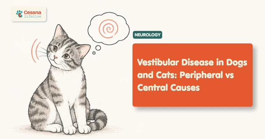
A dog that suddenly can't walk straight, holds its head sharply to one side, and rolls across the floor looks like it's having a stroke. In most older dogs, it isn't. Vestibular disease affects approximately 8 in every 10,000 dogs in primary veterinary practice (VetCompass UK, 2020), and the majority of cases are peripheral, meaning the inner ear rather than the brain is the source. But some aren't, and that distinction matters enormously for treatment, prognosis, and whether advanced imaging is needed right now.
The clinical challenge is that peripheral and central vestibular disease share dramatic, overlapping signs. Both cause a head tilt, ataxia, and abnormal eye movements. What separates them is a set of specific neurological findings that any owner can begin to notice, and that a veterinary neurologist can rapidly clarify.
This guide covers what vestibular disease is in both dogs and cats, how to read the signs, how the peripheral-versus-central distinction is made, and when a neurology consultation with CT or MRI imaging is the right next step.
Key Takeaways
- Vestibular disease affects around 8 in 10,000 dogs, with a median age at diagnosis of 12.68 years (VetCompass UK, 2020).
- Peripheral disease (inner ear/cranial nerve VIII) carries a good-to-excellent prognosis; central disease (brainstem/cerebellum) carries a guarded prognosis and requires urgent imaging.
- A nystagmus rate of 66 beats per minute or higher is 95% specific and 85% sensitive for peripheral vestibular disease (PMC, 2025).
- Most dogs with idiopathic vestibular disease improve within 72 hours and recover fully within 2 to 3 weeks.
Table of Contents
- What Is Vestibular Disease in Dogs and Cats?
- What Are the Signs of Vestibular Disease?
- Head Tilt and the Balance Disorder Every Owner Notices
- Ataxia, Circling, and Falling: What Dog Walking in Circles Really Means
- How Do Vets Tell Peripheral from Central Vestibular Disease?
- What Causes Peripheral Vestibular Disease?
- What Causes Central Vestibular Disease?
- How Is Vestibular Disease Diagnosed? Neurology Consultation and CT Imaging
- Treatment and Recovery: What to Expect
- When Does Vestibular Disease Require Emergency Care?
- Frequently Asked Questions
- Next Steps
What Is Vestibular Disease in Dogs and Cats?
Vestibular disease is an umbrella term for any disorder disrupting the vestibular system, the sensorimotor network that maintains a pet's sense of balance, spatial orientation, and coordinated eye movement. This system has two anatomically distinct components, and the location of the lesion determines the entire diagnostic and treatment pathway.
The peripheral vestibular system consists of the inner ear (semicircular canals and otolith organs), the vestibulocochlear nerve (cranial nerve VIII), and its connections to the brainstem. Disorders here include inner-ear infections, idiopathic degeneration, polyps, and trauma.
The central vestibular system consists of the vestibular nuclei in the brainstem and the flocculonodular lobe of the cerebellum. Disorders here include brain tumors, inflammatory or infectious encephalitis, strokes, and metabolic disease affecting the brain.
Both systems produce strikingly similar outward signs, which is why distinguishing them requires a neurological examination, not just observation from across the room.
In cats, a 2025 study of 196 cases with peripheral vestibular syndrome found that otitis media/interna (OMI) was the most common diagnosis at 71.4%, followed by idiopathic vestibular syndrome at 24%, middle ear neoplasia at 3.6%, and congenital causes at 1% (Veterinary Record, 2025). The pattern in dogs skews differently, with idiopathic vestibular disease dominating in older animals.
canine neuroanatomy overview
What Are the Signs of Vestibular Disease?
Vestibular disease in both dogs and cats produces a recognizable cluster of signs that appear suddenly and can be terrifying to witness. The median age at diagnosis in dogs is 12.68 years (VetCompass UK, 2020), so owners of senior dogs should know these signs before they happen.
Head Tilt and the Balance Disorder Every Owner Notices
Head tilt is the most consistent and visible sign of vestibular disease. The animal holds its head rotated to one side, with the affected ear tilted toward the ground. It's not occasional or subtle; it's persistent and often dramatic.
The tilt reflects an asymmetric signal from the two sides of the vestibular system. When one side is damaged, the brain receives unequal inputs about body position, and the animal tilts toward the damaged side as if being pulled.
Accompanying the head tilt, you'll typically see:
- Nystagmus: involuntary, rhythmic eye movements; the eyes flick back and forth or rotate
- Strabismus: a positional eyeball deviation, one eye drops or drifts downward when the head is lifted
- Nausea and vomiting: the vestibular disruption triggers motion sickness responses
- Wide-based stance or crouching: the animal tries to lower its center of gravity
- Excessive drooling: driven by nausea
These signs can appear within minutes of onset and are typically at their worst in the first 24 to 48 hours.
Ataxia, Circling, and Falling: What Dog Walking in Circles Really Means
Ataxia is the medical term for the incoordination that makes a dog or cat look drunk or disoriented. In vestibular disease, it reflects the brain's inability to interpret spatial position accurately. The animal's limbs move but not in the right directions.
Dog walking in circles is one of the most alarming signs owners describe. The circling is typically toward the side of the lesion: a dog with a left-sided vestibular problem tends to circle left, lean left, and fall left. It's not random wobbling; there's a directional pattern.
Severe cases show rolling: the animal actively rolls across the floor and can't stop itself. This can look like a seizure but isn't one. Rolling and inability to stand are vestibular signs, not motor paralysis.
seizure vs vestibular episode in dogs
How Do Vets Tell Peripheral from Central Vestibular Disease?
Distinguishing peripheral from central vestibular disease is the most clinically important decision in this condition's management: a 2025 PMC study confirmed that a resting nystagmus rate of 66 beats per minute or higher is 95% specific and 85% sensitive for peripheral vestibular disease (PMC, 2025). Peripheral disease has a good-to-excellent prognosis; central disease carries a guarded prognosis and demands urgent advanced imaging.
<figcaption>Sources: PMC12204086 (2025); PMC7249679 (2020); dvm360 vestibular proceedings. BPM = beats per minute of nystagmus.</figcaption> </figure>
The two clinical features that most reliably flag central disease are:
Vertical nystagmus: nystagmus whose fast phase beats upward or downward rather than horizontally. This pattern is essentially never seen in peripheral disease and points directly to a brainstem lesion until proven otherwise.
Postural reaction deficits: if a veterinarian tests the knuckling response (placing the paw on its dorsum) and the animal can't correct it, or if proprioception is impaired, the lesion is almost certainly central. The peripheral vestibular system doesn't directly control limb proprioception; only central lesions do.
Altered mental status is another strong central indicator. A dog with peripheral vestibular disease is alert, responsive, and often quite distressed by its inability to balance. A dog with a brainstem lesion may be obtunded, stuporous, or behaviorally inappropriate.
According to the 2025 penlight-cover test study, measuring nystagmus rate at the bedside takes under two minutes and significantly narrows the differential before any imaging is performed (PMC, 2025). Peripheral nystagmus beats at a median of 90 BPM; central vestibular cases show median nystagmus rates near 0 BPM.
neurological exam in dogs and cats
What Causes Peripheral Vestibular Disease?
Peripheral vestibular disease is far more common than central disease, and in dogs, idiopathic vestibular disease is the single most frequent cause. The 2020 VetCompass UK study confirmed a median onset age of 12.68 years, earning the label "old dog vestibular disease" (VetCompass UK, 2020). The cause is unknown, the onset is sudden, and the resolution is typically spontaneous.
Common peripheral causes in dogs:
- Idiopathic vestibular disease: most common in dogs over 8 years, no identifiable cause, often self-resolving
- Otitis media/interna: bacterial or yeast infection extending from the outer ear canal into the middle and inner ear; often accompanied by head shaking, ear discharge, or facial nerve paralysis
- Hypothyroidism: low thyroid hormone levels can cause peripheral neuropathy affecting CN VIII
- Trauma: blunt head trauma or fracture of the petrous temporal bone
- Nasopharyngeal polyps: more common in cats; polyps originating from the Eustachian tube or tympanic cavity
- Ototoxic medications: aminoglycoside antibiotics (gentamicin, tobramycin), certain ear-cleaning agents
In cats, the 2025 study of 196 peripheral vestibular cases found that otitis media/interna accounted for 71.4% of diagnoses, and affected cats were significantly younger than those with idiopathic disease and more likely to have a prior history of ear or respiratory signs (Veterinary Record, 2025). Cats with idiopathic vestibular syndrome, like their canine counterparts, tend to recover without intervention.
<figcaption>Source: Caldero Carrete et al., Veterinary Record, 2025 (n = 196 cats with peripheral vestibular syndrome).</figcaption> </figure>
ear infections in dogs and cats
What Causes Central Vestibular Disease?
Central vestibular disease is less common but significantly more serious. It involves structures inside the skull (the brainstem and cerebellum) and the underlying causes range from strokes to brain tumors to infectious encephalitis.
Common central causes in dogs and cats:
- Brain tumors: meningioma, choroid plexus tumor, glioma; meningioma is the most common primary brain tumor in both species
- Vascular events (strokes): ischemic or hemorrhagic strokes affecting the brainstem or cerebellar territory; more common in dogs with hypertension, hypothyroidism, or hyperadrenocorticism
- Inflammatory encephalitis: immune-mediated (GME, NME in small breeds), infectious (FIP in cats, distemper, Cryptococcus)
- Thiamine deficiency: particularly in cats on all-fish diets or that have recently stopped eating; causes focal brainstem lesions
- Metronidazole toxicity: high doses of this common antibiotic can cause reversible central vestibular signs in both species
The pattern of cranial nerve deficits in central vestibular disease often involves multiple cranial nerves simultaneously: for example, facial nerve paralysis plus vestibular signs plus a depressed gag reflex, because brainstem lesions affect several cranial nerve nuclei in proximity to each other. This multi-nerve involvement is essentially never seen in pure peripheral vestibular disease.
How Is Vestibular Disease Diagnosed? Neurology Consultation and CT Imaging
The neurological examination is the primary diagnostic tool. A veterinary neurologist evaluates nystagmus character and rate, postural reactions, cranial nerve function, mental status, and gait in a systematic way that takes roughly 15 to 30 minutes. For straightforward peripheral presentations in older dogs, the examination finding alone may be sufficient to make a presumptive diagnosis and begin a trial of supportive care.
When advanced imaging is needed:
| Situation | Appropriate Next Step |
|---|---|
| Classic old-dog vestibular, improves within 72 hours | Monitor; no imaging required |
| Classic old-dog vestibular, no improvement at 72 hours | Bloodwork (thyroid), then CT/MRI |
| Any sign of central disease (vertical nystagmus, postural deficits, altered mentation) | MRI urgent, same day if possible |
| Cat with facial nerve paralysis + vestibular signs | CT of tympanic bullae first; MRI if CT negative |
| Young dog or cat with vestibular signs (no "old dog" profile) | Full workup including imaging |
| Gradual onset or progressive worsening over weeks | MRI regardless of peripheral-appearing signs |
CT vs. MRI in vestibular disease:
CT scanning is the preferred first-line modality for evaluating the middle and inner ear, because it provides excellent bone detail and can identify fluid in the tympanic bulla, bone erosion from otitis, or middle ear masses. CT is faster, cheaper, and requires shorter anesthesia than MRI.
MRI is superior for evaluating the brainstem and cerebellum, which don't image well on CT. Any case with central signs warrants MRI. Both procedures require general anesthesia in dogs and cats, which is why the neurological examination is so valuable as a triage tool: it determines which imaging modality is appropriate and whether imaging is needed at all.
Cerebrospinal fluid (CSF) analysis is often performed at the same anesthetic event as MRI when encephalitis is suspected. It provides definitive information about inflammatory and infectious central causes.
what to expect at a veterinary neurology referral
Treatment and Recovery: What to Expect
Treatment for vestibular disease depends entirely on the underlying cause, which is why the peripheral-versus-central distinction is so clinically urgent.
Idiopathic peripheral vestibular disease requires no specific treatment beyond supportive care. Anti-nausea medication (maropitant or meclizine) reduces vomiting and distress. The pet needs a safe environment: remove access to stairs, pools, and elevated surfaces, and add non-slip flooring to prevent falls. Most dogs begin improving within 72 hours of onset and achieve full or near-full recovery within 2 to 3 weeks. A residual head tilt may persist in some animals for life without affecting quality of life.
Otitis media/interna requires targeted antibiotic or antifungal therapy, often for six to eight weeks, based on culture and sensitivity results from ear cytology or surgery. Surgical drainage (bulla osteotomy) may be needed in chronic or unresponsive cases. Recovery is good when treatment is appropriate and timely.
Central vestibular disease treatment targets the underlying cause: steroids and immunosuppressive drugs for encephalitis, surgery or radiation for accessible tumors, antifungals for Cryptococcus, thiamine for deficiency states, and intensive supportive care for strokes. Prognosis varies widely: a stroke in a young dog from a single vascular event can resolve substantially over weeks; a large brainstem glioma carries a poor outlook regardless of intervention.
<figcaption>Recovery timelines are approximate and depend on severity, patient age, and treatment response. Idiopathic vestibular disease data: VCA Hospitals; otitis data: Veterinary Record 2025.</figcaption> </figure>
supportive nursing care for vestibular pets
When Does Vestibular Disease Require Emergency Care?
Most peripheral vestibular disease is not an emergency, but the initial presentation is dramatic enough that it often looks like one. The practical rule: if the signs are improving within 72 hours, the picture is almost certainly peripheral and benign. If they're worsening or stable without improvement, urgent evaluation is needed.
Go to an emergency veterinarian or veterinary neurologist immediately if:
- The animal shows vertical nystagmus at any point
- Mental status is altered: the pet is unresponsive, stuporous, or behaviorally confused
- Limb weakness or paresis accompanies the vestibular signs
- Multiple cranial nerve deficits are visible (facial drooping + vestibular signs simultaneously)
- Signs are progressing rapidly rather than staying stable or improving
- The animal is a young dog or cat (idiopathic vestibular disease favors older animals; young animals with vestibular signs need a workup)
- No improvement in the first 72 to 96 hours of supportive care
The first 72 hours of watching for improvement is essentially a bedside diagnostic test. Improvement confirms peripheral disease and removes the urgency for imaging. No improvement or worsening confirms the need for advanced imaging and neurology referral.
finding a board-certified veterinary neurologist
Frequently Asked Questions
What does vestibular disease look like in dogs?
Vestibular disease in dogs typically appears suddenly and severely: the dog can't walk straight, holds its head tilted sharply to one side, has rapid involuntary eye movements (nystagmus), may vomit, and sometimes rolls across the floor. Signs are usually worst in the first 24 to 48 hours. Most cases in older dogs are idiopathic, begin improving within 72 hours, and resolve within 2 to 3 weeks without specific treatment.
How do you tell vestibular disease apart from a stroke in dogs?
In dogs, strokes are far less common than idiopathic vestibular disease, but the signs overlap significantly. Central vestibular signs that suggest a stroke include vertical nystagmus, altered mental status, multiple cranial nerve deficits, or limb weakness. A dog that is fully alert, has purely horizontal nystagmus, and is improving within 72 hours almost certainly doesn't have a stroke. MRI is the only definitive way to identify a brainstem or cerebellar stroke.
Is vestibular disease in cats different from dogs?
Yes, in one important way: cats are far more likely to have an infectious cause. A 2025 study of 196 cats found that otitis media/interna accounted for 71.4% of peripheral vestibular cases, compared to the idiopathic category that dominates in dogs (Veterinary Record, 2025). Any cat presenting with vestibular signs, especially one with a history of ear discharge or upper respiratory infections, should have the middle ear evaluated with CT before idiopathic disease is assumed.
Does my pet need a CT or MRI for vestibular disease?
Not always. If the neurological examination shows clear peripheral features (horizontal or rotatory nystagmus, fast rate, intact postural reactions, normal mental status) and the pet begins improving within 72 hours, no imaging is needed. CT of the tympanic bullae is indicated when otitis media/interna is suspected (especially in cats). MRI of the brain is urgent when central signs are present, in any young animal, or when peripheral signs fail to improve by 96 hours.
Can vestibular disease cause permanent damage?
Most peripheral vestibular disease resolves fully. A minority of dogs and cats retain a persistent head tilt that never fully resolves, but this doesn't affect their quality of life significantly. Central vestibular disease can cause permanent deficits depending on the underlying cause and how quickly treatment is started. A vascular event (stroke) may resolve substantially over weeks, while a brain tumor will continue to progress without active management.
How is a neurology consultation different from a regular vet visit?
A veterinary neurologist is a specialist with additional years of residency training in diseases of the nervous system, completing the American College of Veterinary Internal Medicine (ACVIM) neurology board certification. They perform a structured neurological examination that localizes the lesion to a precise region of the nervous system before ordering any tests. For vestibular disease specifically, a neurologist can often determine peripheral vs. central disease at the bedside, avoiding the cost of imaging when it isn't needed, or correctly triaging to urgent imaging when it is.
Next Steps
Vestibular disease in dogs and cats is usually far less catastrophic than it looks in the first hour. The peripheral-versus-central distinction is the pivotal question, and the answer usually comes from the neurological examination, not the imaging suite. A dog that is alert, has horizontal nystagmus, and is improving by the next morning is almost certainly going to be fine within a few weeks.
If signs are worsening, a young animal is affected, or any central features are present (vertical nystagmus, limb weakness, or altered mentation), treat that as urgent. Get a neurology referral and CT or MRI on the same day.
For animals already in recovery: