Recurrent Ear Infections in Pets: Finding the Underlying Cause
Recurrent ear infections in dogs are almost always a symptom of an underlying disease. Learn how vets find the root cause and stop the cycle of chronic otitis.
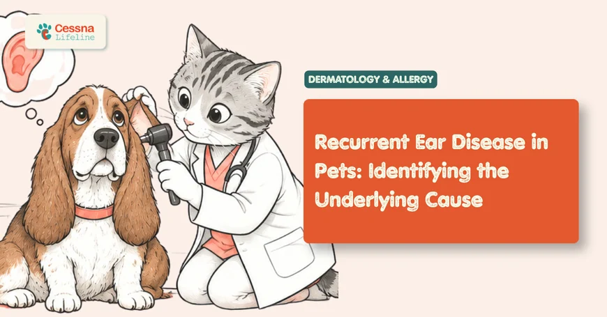
A dog that has been treated for an ear infection three times in twelve months does not have a treatment-resistant ear infection. It has an undiagnosed underlying condition that guarantees each treated infection will return within weeks of completing the antibiotic course. Recurrent otitis externa in dogs is one of the most reliably mismanaged conditions in veterinary medicine, not because the infections are difficult to treat but because they are easy to treat and the apparent cure creates a false endpoint. The infection clears. The underlying cause remains. The next infection begins. dog allergies atopic dermatitis guide
What to Know Recurrent ear infections in dogs and cats are almost never a primary condition. They are symptoms of an underlying disease, most commonly atopic dermatitis, food allergy, or hormonal disease. Treating the infection without identifying the underlying cause produces temporary resolution followed by rapid recurrence. Long-term control requires diagnosing and managing the root cause, not repeating antibiotic courses.
The Ear That Never Fully Heals
The veterinary framework for understanding chronic otitis uses a three-part model: primary causes that initiate the disease, predisposing factors that make the ear canal vulnerable, and perpetuating factors that maintain the infection once established. This model is not academic; it directly determines which interventions will produce lasting results and which will fail on a predictable schedule.
Most dogs presented for a third or fourth course of ear treatment have had the perpetuating factors addressed repeatedly (the yeast or bacteria causing the visible infection) while the primary cause has gone undiagnosed and the predisposing factors have been unmodified. Treating the perpetuating factor alone is not poor medicine in isolation; it is poor medicine in the absence of a plan to identify and address what allowed the infection to establish in the first place.
The otitis triad framework requires the clinician to ask three questions at every recurrence: What underlying disease is generating the abnormal ear environment? What anatomical or behavioral factors are reducing the ear's natural self-cleaning ability? And what organisms are now thriving in that environment, with what resistance profile? Only when all three questions are answered in sequence does treatment have a realistic chance of producing an outcome that does not require repeating the same course of medication in six to eight weeks.
Otitis externa affects approximately 10 to 20% of dogs seen in general veterinary practice, making it one of the most commonly encountered dermatological presentations. In cats, otitis is less common overall but Otodectes cynotis (ear mites) and Malassezia overgrowth are the predominant causes, and the treatment approach differs substantially from the dog.
What Causes Recurrent Ear Infections in Dogs?
Primary causes are the diseases that alter the ear canal environment enough to permit secondary infection. Without a primary cause, a healthy ear canal's anatomy and normal microbiome maintain a self-regulating balance that prevents pathological infection in the vast majority of dogs.
Allergic disease accounts for 80 to 90% of recurrent otitis cases in dogs where a primary cause is identified. Atopic dermatitis is the single most common primary cause; food allergy is the second most common and is clinically indistinguishable from atopic dermatitis on ear examination alone. The allergic ear disease mechanism is direct: IL-4 and IL-13 cytokine signaling in the ear canal skin (same mechanism as skin atopy) disrupts the microbiome balance and increases cerumen production, creating an environment that favors Malassezia and Staphylococcus overgrowth. food allergy in dogs vs. environmental allergy guide
Hypothyroidism is the second most common systemic disease associated with recurrent otitis in middle-aged and older dogs. Thyroid hormone deficiency reduces epidermal cell turnover and sebaceous gland secretion, producing ceruminous accumulation and a dry, hyperkeratotic canal lining that predisposes to secondary infection. Dogs with hypothyroid-associated otitis typically have concurrent signs of thyroid deficiency including weight gain without appetite change, bilateral symmetrical truncal hair loss, and lethargy. hypothyroidism in dogs signs and diagnosis guide
Citation Capsule: Allergic skin disease (atopic dermatitis and food allergy combined) is identified as the primary cause in approximately 80 to 90% of dogs with recurrent otitis externa in which a primary diagnosis is established; Malassezia pachydermatis overgrowth is found in ear canal cytology in more than 50% of atopic dogs, and Staphylococcus pseudintermedius is the most common bacterial pathogen; in dogs with recurrent otitis and no identified parasitic or foreign body cause, dermatological evaluation for underlying allergic disease is the most high-yield diagnostic step rather than further antimicrobial courses (Saridomichelakis et al., 2007). Allergy identification and management is the most consequential intervention for long-term otitis control.
How Do Vets Diagnose the Underlying Cause of Otitis?
Diagnosis of recurrent otitis proceeds in two parallel tracks: identifying the perpetuating organisms to guide the immediate infection treatment, and investigating the underlying primary cause to prevent recurrence.
Ear cytology is the most immediately actionable and underused diagnostic in otitis management. A cotton swab sample from the horizontal ear canal, rolled onto a glass slide, heat-fixed, and stained with a Diff-Quik type stain provides within-appointment identification of the predominant organism type: yeast (Malassezia, recognized by their characteristic peanut-shaped budding morphology), cocci (Staphylococcus), rods (Pseudomonas, Proteus, Klebsiella), or mixed infection. Cytology takes 10 minutes. It determines whether treatment is anti-fungal, antibacterial, or both, and it distinguishes normal commensal yeast counts (under 5 per high-power field) from pathological overgrowth.
Bacterial culture and sensitivity is indicated when: rod organisms are visible on cytology (rod-form bacteria indicate gram-negative infection, most commonly Pseudomonas, which has high multidrug resistance rates); the infection has failed to respond to appropriate initial therapy; or the infection has recurred multiple times with apparently adequate treatment courses. Culture guides antibiotic selection for difficult cases and identifies methicillin-resistant Staphylococcus pseudintermedius (MRSP), which is emerging in recurrent otitis populations.
Otoscopic examination provides assessment of the ear canal architecture and tympanic membrane integrity. Video otoscopy, which projects a magnified image of the canal on a monitor, allows identification of canal wall hyperplasia, polyps, foreign bodies, masses, and tympanic membrane rupture that standard hand-held otoscopes miss in swollen or painful canals. Tympanic membrane rupture changes the treatment approach substantially: ototoxic topical preparations containing gentamicin or chlorhexidine are contraindicated when the membrane is ruptured or integrity is in question.
Primary cause investigation: Any dog with a third episode of otitis that does not have an identified primary cause should receive a systematic dermatological workup. This includes assessment for atopy using the Favrot criteria and, if food allergy is a possibility based on the history, an 8 to 12-week strict elimination diet trial. Thyroid function testing (total T4, free T4, TSH) is appropriate for any middle-aged or older dog with recurrent otitis and no identified allergic cause, particularly in breeds predisposed to hypothyroidism (Golden Retrievers, Doberman Pinschers, Irish Setters, Boxers, Cocker Spaniels).
Citation Capsule: Ear canal cytology correctly identifies the dominant infection type (yeast versus cocci versus rod bacterial) in the vast majority of otitis cases and should be performed at every recheck appointment to monitor treatment response, because the organism distribution can shift during treatment; cytological resolution should precede treatment discontinuation regardless of clinical symptom resolution, as dogs may appear clinically improved before the microbial overgrowth has cleared, and premature discontinuation is a common cause of rapid recurrence (Cole et al., 1998). Cytology is a treatment endpoint indicator, not a one-time diagnostic.
What Is the Role of Allergies in Chronic Ear Disease?
Allergic otitis is not an infection. It is an inflammatory condition in the ear canal skin that creates the conditions for secondary infection. The Malassezia and Staphylococcus that grow in the allergic ear are opportunistic commensals exploiting a compromised microenvironment, not primary pathogens. Treating them without treating the allergy is pharmacologically equivalent to draining a pool that has a running hose filling it. dog allergies atopic dermatitis environmental allergies
The mechanism in the allergic ear follows the same Th2 cytokine pathway as atopic skin disease more broadly. IL-4 and IL-13 signaling in the ear canal epithelium increases ceruminous gland secretion, shifts the fatty acid composition of the cerumen toward a profile that favors Malassezia growth, and impairs the epidermal barrier function that normally limits colonization. The result is an ear canal that perpetually generates conditions favorable to microbial overgrowth even when topical treatment temporarily reduces populations.
In food-allergic dogs, the ear canal may be the only or predominant site of allergic manifestation. Dogs with ear disease as the sole presenting complaint of food allergy account for a clinically significant proportion of food allergy cases, and these dogs may have no gastrointestinal signs, no other cutaneous sites of pruritus, and no seasonal pattern to guide the diagnostic approach. The elimination diet trial remains the only reliable method to diagnose or exclude food allergy in these patients; serum food allergy panels have insufficient sensitivity and specificity to replace the dietary trial.
The practical implication of allergic otitis management is that the antimicrobial component of treatment is always secondary to allergy control. In a dog with well-controlled atopy on lokivetmab or oclacitinib, Malassezia populations in the ear canal often normalize without topical antifungal treatment simply because the allergic microenvironment driving overgrowth has been reduced. In a dog whose atopy is poorly controlled, Malassezia will repopulate within weeks of any topical antifungal course regardless of the agent used.
Citation Capsule: Food allergy presents with otitis as the primary or sole clinical sign in a clinically significant proportion of food-allergic dogs, without concurrent gastrointestinal disease or pruritus at other body sites; these patients are frequently misdiagnosed as having primary recurrent otitis, and repeated antibiotic and antifungal courses without an elimination diet trial fail to achieve remission; resolution of otitis following a strict 8 to 12-week elimination diet trial is both diagnostic and therapeutic, confirming food allergy as the primary cause and demonstrating that allergen elimination alone can control the ear disease without ongoing topical treatment (Paterson, 1995). Food allergy identification eliminates the need for lifelong topical ear treatment in responding dogs.
Which Ear Cleaning Practices Actually Help?
Ear cleaning has an evidence base in chronic otitis management, and not every cleaning approach is equivalent. The clinical value of cleaning depends on what it accomplishes mechanically: removing excess cerumen and exudate reduces the substrate available for microbial overgrowth, improves penetration of subsequently applied topical medications, and maintains the ear canal's natural migration of epithelial cells toward the opening of the canal.
Ceruminolytic cleaners (containing squalene, propylene glycol, or dioctyl sodium succinate) dissolve and emulsify wax accumulations and are appropriate for maintenance cleaning in dogs with ceruminous debris. They should not be left in the canal for extended periods; a 5 to 10-minute dwell time followed by cotton ball absorption at the canal opening is the standard protocol. dog ear cleaning how-to guide
Acidifying cleaners (containing acetic acid, boric acid, or salicylic acid) lower canal pH, which inhibits bacterial and yeast growth directly and makes the canal less hospitable for recurrence. These are appropriate for maintenance use in dogs in remission, particularly those with allergic disease where microenvironmental control between flares can extend the infection-free interval.
Hydrophobic cleaners (containing isopropyl alcohol) dry the canal and are appropriate for dogs whose primary predisposing factor is excessive moisture from swimming. They should not be used in ulcerated canals, which is a contraindication for any alcohol-containing preparation.
The most important ear cleaning scenario in recurrent otitis management is not the owner's home maintenance cleaning but the video-otoscope-guided flush performed under general anesthesia when biofilm is established. Pseudomonas aeruginosa and Staphylococcus pseudintermedius both form robust biofilms on the epithelium of chronically infected ear canals. Biofilm-embedded bacteria are protected from antibiotic penetration by a polysaccharide matrix that reduces effective drug concentration at the bacterial surface by a factor of 100 to 1,000 relative to planktonic bacteria. A Pseudomonas isolate embedded in biofilm may require drug concentrations that no topical preparation can achieve without first mechanically disrupting the biofilm itself. Video-otoscope-guided flushing under general anesthesia with a saline irrigation system physically removes biofilm before topical medication is applied, converting a pharmacologically impossible treatment situation into a manageable one. Owners who are told their dog's ear infection is "antibiotic-resistant" often have a dog with intact biofilm, not intrinsic drug resistance.
Citation Capsule: Bacterial biofilm in chronic canine otitis is documented primarily with Pseudomonas aeruginosa and Staphylococcus pseudintermedius; biofilm-protected bacteria require drug concentrations 100 to 1,000 times higher than planktonic minimum inhibitory concentrations for effective killing, which is achievable with high-concentration topical preparations after biofilm disruption but not through routine topical application to an intact biofilm surface; video-otoscope-guided deep canal flushing under general anesthesia is the required first step before topical antimicrobial therapy in ears with established biofilm, because medication applied to intact biofilm provides minimal antimicrobial efficacy regardless of agent selection (Nuttall, 2016). Biofilm disruption before topical treatment is not optional in Pseudomonas chronic otitis; it determines whether treatment succeeds.
When Is Surgery the Answer for Chronic Ear Disease?
A subset of dogs with long-standing otitis develops irreversible structural changes that make medical management physiologically impossible regardless of the underlying cause being controlled. Canal wall hyperplasia, ulceration, fibrosis, and calcification eventually produce an ear canal that is so anatomically compromised that topical medications cannot penetrate to the depths of infection, the canal cannot self-clean, and medical treatment at any intensity fails to achieve remission. video otoscopy dogs ear disease guide
Lateral wall resection (LWR) removes the lateral wall of the vertical ear canal, creating a wider opening that improves ventilation, drainage, and topical medication access. It is appropriate for dogs with mild to moderate canal hyperplasia in which the horizontal canal remains functional. It does not eliminate the primary cause and requires ongoing medical management.
Total ear canal ablation (TECA) with lateral bulla osteotomy (LBO) removes the entire external ear canal, the tympanic membrane, and the contents of the tympanic bulla. It is the definitive surgical option for end-stage otitis with calcification or severe proliferative disease. Success rates in appropriately selected cases exceed 85 to 90%, with pain elimination as the consistent and immediate outcome. The primary risk is facial nerve paresis (from proximity of the facial nerve to the surgical field), which occurs in approximately 10 to 25% of cases and is usually temporary.
TECA-LBO is not a failure of medical management; it is the correct treatment for a disease that has progressed to the point where medical management is no longer physiologically feasible. Delaying TECA in a dog with calcified, end-stage otitis prolongs chronic pain and does not improve surgical outcomes. The decision is ideally made by or in consultation with a board-certified veterinary surgeon.
The Treatment Strategies That Break the Otitis Cycle
Effective long-term otitis management addresses all three tiers of the otitis triad simultaneously, not sequentially.
Otitis Management: Perpetuating Factor Treatment Guide
| Organism / Change | Clinical Sign | Topical Treatment | Systemic Treatment |
|---|---|---|---|
| Malassezia (yeast) | Dark waxy discharge, musty odor, mild pruritus | Miconazole, clotrimazole, nystatin combination | Oral ketoconazole (severe cases only) |
| Staphylococcus (cocci) | Creamy yellow discharge, pruritus, pain | Neomycin, polymyxin B, fusidic acid | Amoxicillin-clavulanate (sensitive strains) |
| Pseudomonas (rods, MDR) | Green/grey discharge, severe pain, odor | High-concentration tobramycin or enrofloxacin topical | Guided by C&S; systemic often insufficient |
| MRSP | Recurrent Staph, failed multiple courses | Mupirocin topical, silver-based preparations | C&S guided only; biofilm disruption first |
| Canal hyperplasia | Stenosis, reduced medication penetration | Anti-inflammatory (triamcinolone) topical | Oral prednisolone short course |
| Calcification | Palpable firmness, radiographic change | None effective | Surgical (TECA-LBO) |
What veterinary dermatologists observe consistently: Pseudomonas culture sensitivity reports create significant clinical confusion in recurrent otitis management. When a culture report lists Pseudomonas as "resistant" to enrofloxacin by disc diffusion, it means the systemic plasma concentration of enrofloxacin achievable at standard dosing is below the MIC. It does not mean that enrofloxacin-based topical preparations are ineffective. High-concentration topical enrofloxacin formulations (such as Baytril Otic) achieve drug concentrations in the ear canal that are 100 to 1,000 times higher than systemic plasma concentrations, which routinely exceed the MIC of isolates reported as "resistant" on standard culture sensitivity interpretation. The critical distinction is topical versus systemic route, not the drug itself. A Pseudomonas isolate reported as resistant to enrofloxacin should prompt a topical high-concentration preparation, biofilm disruption by deep canal flush, and cytological monitoring, not automatic escalation to systemic aminoglycosides or carbapenems.
Immunological treatment: In allergic otitis, the single most impactful long-term intervention is effective allergy management. Dogs on lokivetmab that achieve significant whole-body pruritus reduction typically show proportional improvement in ear disease frequency and severity. The ear is a cutaneous site; what controls cutaneous atopy also controls aural atopy. Dogs with well-managed atopy who require ear treatment may benefit from maintenance ear cleaning with an acidifying solution two to three times weekly between active infections rather than intermittent treatment of established infections.
Monitoring protocol: Cytological resolution of organisms, not symptom resolution, is the appropriate endpoint for active infection treatment. Dogs treated for 14 to 21 days with appropriate topical therapy may appear clinically improved (reduced pruritus, less discharge) before microbial populations have cleared. Rechecking cytology at the end of every treatment course and continuing treatment until cytology shows fewer than 5 Malassezia per high-power field and no bacteria prevents the "apparent cure" cycle that produces recurrent infections.
Frequently Asked Questions
Why does my dog keep getting ear infections?
Recurrent ear infections almost always indicate an unidentified underlying condition, most commonly atopic dermatitis or food allergy in dogs under 7 years old, or hypothyroidism in older dogs. Each completed antibiotic course treats the secondary infection; the primary condition persisting in the ear canal microenvironment recruits a new infection within weeks. Long-term resolution requires diagnosing and managing the underlying disease, not repeating the same antibiotic course.
What does Pseudomonas ear infection in dogs mean?
Pseudomonas aeruginosa in the ear canal indicates a chronic, deep infection with a gram-negative rod that is frequently multidrug-resistant. Its presence suggests the infection has been present for a significant time and has progressed beyond what Staphylococcus and Malassezia typically represent. Pseudomonas otitis requires culture and sensitivity testing, deep canal flushing under anesthesia to disrupt biofilm, and high-concentration topical antimicrobial therapy. It is not a treatment failure; it is a signal that a more intensive diagnostic and treatment protocol is required.
Can I clean my dog's ears too much?
Yes. Overcleaning with irritating solutions in an inflamed or ulcerated canal can disrupt the normal cerumen and epithelial migration that the ear relies on for self-cleaning. In ears that are actively infected or ulcerated, cleaning should be directed by a veterinarian. In healthy ears between infections, cleaning two to three times per week with an appropriate ceruminolytic or acidifying solution is appropriate for dogs with chronic allergic ear disease. More frequent cleaning than this does not provide additional benefit and may irritate the canal.
Is there a food that causes ear infections in dogs?
No single food causes ear infections universally. In food-allergic dogs, a specific dietary protein (most commonly beef, chicken, dairy, or wheat, depending on the individual dog's sensitization history) triggers an immune-mediated allergic response in the ear canal skin that creates conditions for secondary Malassezia or bacterial overgrowth. Identifying which protein the dog is sensitized to requires an 8 to 12-week elimination diet with a hydrolyzed or novel protein diet, not commercial "hypoallergenic" food with multiple protein sources.
When should I see a veterinary dermatologist for my dog's ear?
Any dog with a third ear infection in twelve months, an ear infection involving rod bacteria (Pseudomonas) on cytology, an ear infection that has not resolved after two appropriate treatment courses, or an ear infection in which the tympanic membrane is ruptured should be evaluated by or in consultation with a veterinary dermatologist. Dermatologists have access to video otoscopy, deep canal flushing capability, allergy testing, and surgical consultation pathways that general practitioners may not.
The Ear That Heals When the Cause Is Found
The frustration that owners bring to a fourth or fifth ear treatment appointment is not about the infection. It is about the cycle: clean, treat, resolve, return. That cycle is predictable when the underlying cause is untreated and permanent when the underlying cause is identified and managed. An atopic dog whose allergy is controlled with monthly Cytopoint injections does not reliably stop getting ear infections because of the Cytopoint; it stops because the allergic microenvironment that made the ear a hospitable host for Malassezia no longer exists at the same intensity. The ear cleaned itself. That outcome is available to most dogs with recurrent otitis, and it begins with asking the question that drives the entire framework: what underlying condition made this ear a place where infections could not stop returning?