Autoimmune Disease in Dogs: When the Body Attacks Itself
In dogs with IMHA, 31% died within 3 months in a 2024 study. Learn about IMHA, lupus, pemphigus, and how vets diagnose and treat immune-mediated disease.
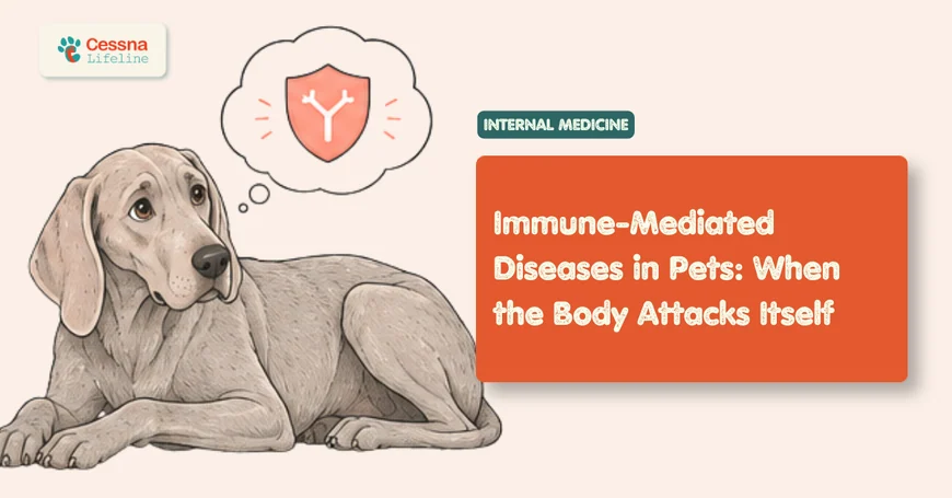
The immune system is supposed to be the body's protector: a precisely calibrated network of cells, proteins, and signals that identifies threats and eliminates them. When it works correctly, a dog recovers from infections, heals wounds, and tolerates its own healthy tissue without incident. When it doesn't, the consequences can be swift, severe, and confusing for everyone involved.
Autoimmune disease in dogs, and the broader category of immune-mediated conditions, occurs when that protective system loses the ability to distinguish between invaders and the body's own cells. It mounts an attack on itself. Red blood cells, platelets, joints, skin, and kidneys can all become targets. The result ranges from manageable with ongoing treatment to immediately life-threatening, depending on which tissue is under attack and how quickly the disease is recognized.
For dog owners, this category of disease is particularly disorienting. A pet can deteriorate rapidly with no obvious cause. The symptoms often mimic infections or other common illnesses. And the diagnosis requires ruling out a long list of other possibilities before a pattern emerges. Understanding what immune-mediated diseases are, how they present, and what modern veterinary medicine can do for them changes the conversation from panic to informed action.
veterinary internal medicine specialists
What to Know Autoimmune disease in dogs occurs when the immune system attacks the body's own healthy tissue, causing conditions like IMHA, IMTP, pemphigus, and lupus. In a 2024 retrospective study of 104 dogs with immune-mediated hemolytic anemia, mortality reached 31% at three months, underscoring that early diagnosis and aggressive treatment are not optional.
What Is Autoimmune Disease in Dogs?
Autoimmune disease is not a single condition. It's a category of diseases united by a common mechanism: the immune system's failure to recognize self from non-self. Normally, a process called central and peripheral tolerance teaches immune cells to leave the body's own tissues alone. When those tolerance mechanisms break down, the immune system produces antibodies or deploys T-cells against the dog's own healthy cells as if they were pathogens.
In dogs, immune-mediated diseases are considered uncommon as a group, though several specific conditions, particularly immune-mediated hemolytic anemia, immune-mediated thrombocytopenia, and pemphigus foliaceus, occur with enough frequency to make them familiar presentations in internal medicine and emergency practice (VCA Animal Hospitals, 2024). They are more common in unspayed females across most conditions, and certain purebred dogs carry documented genetic predispositions.
The immune system's attack can be primary, meaning no identifiable trigger is found, or secondary, triggered by an underlying infection, cancer, drug exposure, or another disease. This distinction matters diagnostically and therapeutically. A dog with secondary immune-mediated disease whose underlying trigger is left untreated will not respond well to immunosuppression alone. Finding the trigger, when one exists, is part of the workup.
What we've found in practice: Secondary immune-mediated disease is consistently underappreciated at first presentation. A dog with IMHA who has a tick-borne disease as the trigger, or a dog with IMTP secondary to heartworm disease, may be managed as primary autoimmune disease for weeks before the underlying condition is identified. A thorough infectious disease panel at the time of first diagnosis is not optional.
How Does the Immune System Turn Against Itself?
The mechanisms vary by disease type, but a few pathways account for most immune-mediated conditions in dogs.
In the case of immune-mediated hemolytic anemia and thrombocytopenia, the immune system produces antibodies that bind to the surface of red blood cells or platelets, tagging them for destruction. Complement proteins are activated. Cells coated in antibody are recognized by macrophages in the spleen and liver as foreign and phagocytosed. The resulting destruction of red blood cells produces profound anemia; the destruction of platelets produces life-threatening bleeding risk.
In inflammatory joint diseases and systemic lupus, immune complexes, aggregates of antibodies and antigens, deposit in tissues including joint capsules, kidneys, and blood vessel walls. The deposition triggers intense local inflammation that damages the tissue over time, even though the initial immune response wasn't targeting that tissue specifically.
In pemphigus, the immune system produces antibodies against the proteins that hold skin cells together. The result is a loss of cell-to-cell adhesion in the epidermis, causing the characteristic blistering and crusting that defines the disease. The same mechanism, targeting different proteins, produces the different forms of pemphigus seen in dogs.
What triggers the initial breakdown of tolerance? That question doesn't have a clean answer. Genetics plays a significant role, particularly in breeds that show clustering of immune-mediated diseases. Environmental factors including UV radiation, certain medications (most notably sulfonamide antibiotics), infections, and possibly vaccinations in genetically susceptible animals have all been proposed as triggers. In most cases of primary disease, no single identifiable trigger is found.
<figcaption>The three major immune mechanisms behind autoimmune diseases in dogs determine which tissues are affected and what clinical signs appear.</figcaption> </figure>
What Are the Most Common Immune-Mediated Diseases in Dogs?
Several distinct conditions fall under the immune-mediated umbrella. They vary significantly in their target tissue, severity, and prognosis, and they're worth understanding individually rather than as an undifferentiated group.
Immune-Mediated Hemolytic Anemia (IMHA)
IMHA is among the most serious immune-mediated conditions a dog can develop. The immune system produces antibodies against the dog's own red blood cells, which are then rapidly destroyed in the spleen and liver. The resulting anemia can be severe enough to be immediately life-threatening.
The statistics are sobering. A 2024 retrospective study of 104 dogs with IMHA found mortality rates of 16% at one month and 31% at three months (Irish Veterinary Journal, 2024). Most deaths occur within the first two weeks of hospitalization, when immunosuppressive medications have not yet reached full effect and the risk of thromboembolic complications is highest (MSPCA-Angell, 2025). Those who survive the acute phase face a relapse rate of 6 to 13%.
Clinical signs reflect the severity of anemia: pale or yellow (icteric) gums, extreme fatigue, rapid breathing, weakness, and in some cases, dark red or brownish urine from hemoglobin released by destroyed red blood cells. The diagnosis typically involves a positive Coombs test confirming antibody coating on red blood cells, combined with spherocytes visible on a blood smear.
Treatment requires aggressive immunosuppression, typically starting with high-dose corticosteroids, often combined with additional agents like azathioprine, cyclosporine, or mycophenolate. Many dogs require blood transfusions to stabilize them during the critical early window. The risk of blood clots is significant enough that anticoagulant therapy is often added.
IMHA affects multiple breeds, but Cocker Spaniels, English Springer Spaniels, and Old English Sheepdogs show higher reported incidence. Middle-aged dogs are most commonly affected, and females may be overrepresented in primary disease.
Immune-Mediated Thrombocytopenia (IMTP)
Where IMHA targets red blood cells, IMTP targets platelets. Antibodies bind to platelet surfaces, causing them to be destroyed by the spleen before they complete their normal lifespan of about 5 to 10 days. Platelet counts can fall to levels where spontaneous hemorrhage becomes the immediate concern.
The presenting picture is often striking: dogs with IMTP commonly show petechiae (tiny pinpoint hemorrhages) or ecchymoses (larger bruising) on the skin and mucous membranes, which appear in 66% of dogs on physical examination (JAVMA, 2011). Melena, the passage of dark tarry stools indicating bleeding in the upper GI tract, occurs in about 25% of cases. Lethargy and reduced appetite are the most common owner-reported complaints at 46% and 34% respectively.
The prognosis for primary IMTP is considerably better than for IMHA. In a study of 73 dogs with primary ITP, 84% survived to hospital discharge (AVMA Journal, 2011). The principal challenge is relapse: 39% of surviving dogs experienced a recurrence. Treatment follows the same immunosuppressive approach used for IMHA, with platelet transfusions reserved for dogs with active life-threatening hemorrhage.
Pemphigus Foliaceus
Pemphigus foliaceus (PF) is the most common autoimmune skin disease in dogs and accounts for roughly one-third of all canine autoimmune conditions (VCA Animal Hospitals, 2024). Unlike the hematologic conditions above, PF's primary target is the skin, specifically the proteins that hold skin cells together in the outermost layer.
The disease presents as pustules, crusts, scaling, and erosions, typically starting on the face, ears, and footpads before spreading more broadly. The footpad involvement is particularly characteristic. Unlike many infectious skin conditions, PF tends to be bilaterally symmetrical, which is a useful early diagnostic clue.
Recent outcomes data is more encouraging than historical reports. A study of 41 dogs with PF found that 52% achieved complete remission, 35% achieved partial remission, and only 13% were ultimately euthanized (PubMed, 2021). Treatment requires long-term immunosuppression, and most dogs will need medication for life, though the dose can often be reduced to the minimum needed to maintain control.
Akita Inus and Chow Chows carry documented breed predispositions. The disease can be triggered or exacerbated by sun exposure, which is why UV protection is sometimes recommended for affected dogs.
Systemic Lupus Erythematosus (SLE)
SLE is often called "the great imitator" in veterinary medicine, and for good reason. It can simultaneously affect the skin, joints, kidneys, blood cells, nervous system, and heart, producing a constellation of symptoms that rarely points immediately to a single diagnosis.
The signature of canine SLE in published case series is consistent: fever appears in 100% of dogs, non-erosive polyarthritis in 90.6%, and kidney involvement in 65.3% (ScienceDirect / Canine Systemic Lupus Erythematosus, 2024). Skin lesions, anemia, and neurological signs can all appear and may dominate the clinical picture at different points in the disease course.
Diagnosis is challenging precisely because no single test confirms SLE. Most veterinary internists use a criteria-based approach adapted from human rheumatology, requiring at least two to three consistent findings. Afghan Hounds, Beagles, German Shepherds, Collies, Poodles, Nova Scotia Duck Tolling Retrievers, and Shetland Sheepdogs are among the breeds with documented predispositions (VCA Animal Hospitals, 2024).
Immune-Mediated Polyarthritis
Immune-mediated polyarthritis (IMPA) occurs when immune complexes deposit in joint capsules, triggering painful inflammation in multiple joints simultaneously. The hallmark is shifting lameness, where the affected leg changes from day to day, combined with systemic signs like fever and lethargy. Unlike infectious arthritis or degenerative joint disease, IMPA is not erosive in most cases, meaning joint cartilage and bone are not destroyed.
A 2024 UK review of 84 dogs with IMPA found that the most commonly affected breeds included cross breeds, Cocker Spaniels, Labrador Retrievers, Border Collies, and Springer Spaniels (Veterinary Medicine and Science / Wiley, 2024). Response to immunosuppressive therapy is generally good, though relapse is a meaningful concern across all nonerosive forms of the disease.
What Are the Symptoms of Autoimmune Disease in Dogs?
One of the most challenging aspects of this disease category is that there's no single symptom that says "autoimmune." The signs depend entirely on what tissue is being attacked. A dog with IMHA looks pale and exhausted; a dog with pemphigus has a crusting face and painful paws; a dog with IMPA is stiff and feverish. They don't look like they have the same underlying mechanism.
What they do share is a quality of being disproportionate to what common explanations would suggest. The anemia is severe. The joint pain affects multiple limbs without a clear injury. The skin lesions don't respond to antibiotics. The dog isn't improving despite treatment for what looks like an infection.
In our experience: Owners of dogs with immune-mediated disease often describe a sense that "something is really wrong" before they can articulate exactly what. The dog is different in a way that goes beyond ordinary illness. That perception is clinically useful. When an owner describes progressive deterioration that doesn't match a simple explanation, the immune-mediated differential deserves serious consideration.
The symptoms worth tracking across all immune-mediated conditions include:
- Pale, white, or yellow-tinged gums (anemia or icterus from IMHA)
- Unexplained bruising, petechiae, or bleeding from the nose or gums (IMTP)
- Shifting lameness that doesn't correspond to a specific injury
- Persistent or recurring fever without an infectious source
- Skin lesions that are bilaterally symmetrical, crusting, or unresponsive to antibiotics
- Extreme fatigue disproportionate to other findings
- Dark urine (hemoglobinuria from severe IMHA)
<figcaption>Frequency of key clinical findings in dogs presenting with immune-mediated thrombocytopenia. Petechiae and bruising are the most consistent physical exam findings.</figcaption> </figure>
No single symptom is diagnostic. The pattern, the combination, the progression, and the failure to respond to simpler treatments are what point toward an immune-mediated process.
recognizing pale gums in dogs
<figcaption>Survival and remission rates vary significantly across immune-mediated diseases. IMHA carries the highest acute mortality risk of the three.</figcaption> </figure>
How Do Veterinarians Diagnose Immune Disorders?
Diagnosing immune-mediated disease is partly a matter of finding consistent abnormalities and partly a matter of ruling out the many conditions that can mimic them. There's rarely a single test that closes the case.
The workup for a dog suspected of having an immune-mediated disease typically begins with a minimum database: complete blood count, serum chemistry panel, and urinalysis. For dogs with IMHA, the blood count shows profound anemia and the smear reveals spherocytes, misshapen red blood cells that result from partial phagocytosis. A direct Coombs test, which detects antibodies coating red blood cells, confirms the immune-mediated nature of the anemia in most cases.
For IMTP, a platelet count below 30,000 per microliter combined with clinical bleeding is highly suggestive. Bone marrow examination may be indicated to confirm that platelet production is adequate, ruling out disease of the marrow itself.
| Condition | Key Diagnostic Test | Supporting Findings |
|---|---|---|
| IMHA | Direct Coombs test positive; spherocytes on smear | Severe anemia, elevated bilirubin, positive autoagglutination |
| IMTP | Platelet count < 30,000/μL; bone marrow evaluation | Petechiae, ecchymosis, melena |
| Pemphigus Foliaceus | Skin biopsy with histopathology | Acantholytic cells, pustule pattern |
| SLE | Criteria-based diagnosis (2–3 of 11 criteria) | ANA titer, protein in urine, polyarthritis, anemia |
| IMPA | Joint fluid cytology from multiple joints | Neutrophilic inflammation, no bacteria |
For SLE and IMPA, joint fluid analysis is central to the diagnosis: a fluid sample showing non-infectious, neutrophil-rich inflammation from at least two joints is the minimum needed to confirm IMPA. Antinuclear antibody (ANA) titers, while not specific to SLE alone, support the diagnosis when positive alongside other criteria.
A consistency worth noting: Infectious disease testing should be standard at the time of first workup for any immune-mediated disease, not added as an afterthought if the case doesn't respond. Ehrlichia, Anaplasma, Babesia, and Leishmania can all cause clinical pictures indistinguishable from primary immune-mediated disease, and treating with immunosuppression without controlling the infection first can be catastrophic.
tick-borne disease testing in dogs
What Does Treatment Look Like?
Across most immune-mediated diseases in dogs, treatment follows the same foundational logic: suppress the immune response enough to stop the destruction, while managing the organ damage that's already occurred and preventing complications. The challenge is that the drugs used to suppress the immune system also reduce resistance to infection, so the treatment itself creates new vulnerabilities.
Glucocorticoids, principally prednisone or dexamethasone, are almost always the starting point. They work quickly, reduce inflammation, and in mild cases may be sufficient on their own. For more severe or refractory cases, secondary immunosuppressives are added: azathioprine, cyclosporine, mycophenolate mofetil, chlorambucil, and leflunomide are all used in dogs, often in combination tailored to the individual patient's disease severity and comorbidities.
For IMHA specifically, the thromboembolism risk demands additional management. Aspirin at anti-platelet doses, heparin, or low-molecular-weight heparins are commonly added. Desmopressin and other supportive therapies may be used in critical cases.
The duration of treatment is another challenge. Most dogs with primary immune-mediated disease require months of medication, with gradual tapering once remission is achieved. For pemphigus and some forms of SLE, lifelong treatment at the lowest effective dose is the realistic goal.
The tapering question: One of the most common sources of relapse in IMHA and IMTP is premature reduction of immunosuppression. Owners understandably want to reduce the medications; the side effects of long-term steroids are real, including weight gain, muscle loss, increased thirst, and susceptibility to infection. But clinical recovery doesn't equal immunologic recovery, and tapering too quickly before the underlying immune dysregulation has settled is a well-documented cause of relapse. The schedule should be driven by objective parameters, particularly repeat CBC values, not by how well the dog appears to be doing.
Frequently Asked Questions
What are the most common autoimmune diseases in dogs?
The most common immune-mediated diseases in dogs are immune-mediated hemolytic anemia (IMHA), immune-mediated thrombocytopenia (IMTP), pemphigus foliaceus, systemic lupus erythematosus (SLE), and immune-mediated polyarthritis (IMPA). Pemphigus foliaceus is the most frequent autoimmune skin disease and accounts for roughly one-third of all canine autoimmune conditions (VCA Animal Hospitals, 2024).
What are the symptoms of autoimmune disease in dogs?
Symptoms vary by disease type. IMHA causes pale or yellow gums, extreme fatigue, and dark urine. IMTP causes bruising, petechiae, and bleeding from body openings. Pemphigus produces crusty, painful skin lesions on the face and footpads. SLE can cause fever, joint pain, anemia, and kidney disease simultaneously. The unifying feature is that symptoms seem disproportionate to common explanations and often fail to respond to routine treatment.
How is IMHA diagnosed in dogs?
IMHA is diagnosed through a combination of findings: severe anemia on bloodwork, spherocytes visible on a blood smear, positive autoagglutination (red blood cells clumping), and a positive Coombs test confirming antibody coating on red blood cells. Supporting tests include elevated bilirubin and the exclusion of other causes of anemia. A 2024 study of 104 dogs documented 31% three-month mortality, highlighting the urgency of early diagnosis (Irish Veterinary Journal, 2024).
Can autoimmune disease in dogs be cured?
Some dogs with primary immune-mediated disease achieve lasting remission after a course of treatment and can eventually discontinue medication. However, most dogs with pemphigus, SLE, and chronic relapsing IMHA or IMTP require ongoing management, sometimes for life. The goal is control, not cure: the lowest effective dose of immunosuppression that keeps the disease in remission while minimizing side effects.
Which dog breeds are most prone to autoimmune diseases?
Several breeds carry documented predispositions. Cocker Spaniels and English Springer Spaniels are overrepresented in IMHA. Afghan Hounds, German Shepherds, Collies, and Nova Scotia Duck Tolling Retrievers have higher rates of SLE. Akita Inus and Chow Chows are predisposed to pemphigus foliaceus. Nova Scotia Duck Tolling Retrievers also carry known risk for immune-mediated polyarthritis. Unspayed females are overrepresented across most conditions (VCA Animal Hospitals, 2024).
breed-specific health risks
What These Diseases Ask of Us
Immune-mediated disease sits at the intersection of immunology, emergency medicine, and long-term management in a way that few other conditions do. A dog with IMHA may need blood transfusions and intensive care in the first week, ongoing immunosuppression for months afterward, and regular monitoring for the rest of its life. The same is true, in different proportions, for IMTP, pemphigus, and SLE.
What makes these diseases particularly demanding for owners is that the treatment is often as difficult as the disease itself. Immunosuppressive drugs cause real side effects. Monitoring requires regular veterinary visits and bloodwork even when the dog seems fine. And the threat of relapse never fully disappears for dogs with a history of IMHA or IMTP.
The data is not entirely discouraging. Dogs that survive the acute phase of IMHA have reasonable long-term prognoses. The majority of dogs with IMTP survive to discharge. Pemphigus, with appropriate treatment, allows most dogs to live comfortable lives. The window that changes outcomes is the early one, when diagnosis is confirmed quickly and treatment is started aggressively before organ damage accumulates.
These diseases ask something specific of the people who love affected dogs: vigilance, commitment to monitoring, and a willingness to engage with complexity rather than wait for symptoms to resolve on their own. That combination, more than any single medication, is what moves these animals from crisis to stability.