Fever of Unknown Origin in Pets: A Systematic Diagnostic Approach
Fever of unknown origin in dogs requires systematic investigation beyond initial bloodwork. Learn the diagnostic steps vets use when fever does not resolve.
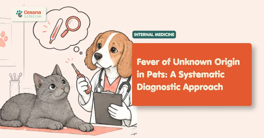
A dog presented for the third time in four weeks with a rectal temperature of 40.1°C, lethargy, and reduced appetite does not have a mystery illness. It has an illness that has not yet been investigated systematically. Fever of unknown origin (FUO) is not a disease; it is a clinical finding that has outrun its initial diagnostic workup. The most common reason a fever remains "unknown" in a dog or cat is not that the cause is inherently undetectable but that the investigation has been reactive rather than systematic: one test at a time, ordered after the previous result fails to yield a diagnosis, with empirical treatment trials obscuring the clinical picture between rounds. The systematic approach replaces this with a pre-specified sequence of investigations that collectively capture the causes responsible for more than 85% of FUO cases. tick-borne diseases in dogs Ehrlichia Anaplasma guide
What to Know Fever of unknown origin in dogs and cats is persistent fever lasting more than 3 weeks without an identified cause after initial diagnostic evaluation. The most common causes are infectious disease (40 to 50%), immune-mediated disease (20 to 30%), and neoplasia (10 to 15%). Systematic staged investigation is required. Administering corticosteroids before infectious causes are excluded is the most consequential diagnostic error in FUO management.
When Fever Is the Clue, Not the Diagnosis
Fever represents a regulated elevation of the hypothalamic thermostat set point in response to endogenous pyrogens, primarily interleukin-1 (IL-1), interleukin-6 (IL-6), and tumor necrosis factor-alpha (TNF-alpha). These cytokines are released by macrophages and monocytes in response to pathogens, immune complexes, tissue necrosis, or neoplastic cell products. The fever itself is a beneficial physiological response: it impairs the replication of temperature-sensitive pathogens and enhances phagocytic function. Suppressing fever with NSAIDs or corticosteroids in a diagnostically unresolved patient reduces the very signal being used to track whether treatment is working.
Normal rectal temperature in dogs ranges from 38.3 to 39.2 degrees Celsius (101 to 102.5 degrees Fahrenheit). Temperatures above 39.4°C (103°F) are elevated; temperatures above 40°C (104°F) constitute significant fever warranting active investigation; temperatures above 41.1°C (106°F) represent hyperthermia or severe fever where direct organ damage becomes a risk and requires immediate intervention regardless of cause.
Hyperthermia (heat stroke, excessive exercise, pain-induced temperature elevation) must be distinguished from fever before the FUO workup begins. Hyperthermia reflects a failure of heat dissipation against an external thermal challenge; the thermostat is set normally but the body cannot shed heat fast enough. A dog that has been confined in a hot car, exercised heavily, or is showing extreme physiological stress from pain may have a rectal temperature above 40°C without true fever. Distinguishing these requires a careful history of events preceding the temperature measurement.
FUO in veterinary medicine is conventionally defined as fever documented on multiple measurements over a minimum of 2 to 3 weeks that has not yielded a diagnosis after initial laboratory evaluation (complete blood count, serum chemistry panel, and urinalysis). The 3-week threshold distinguishes FUO from acute self-limiting febrile illnesses, which typically resolve within 1 to 2 weeks with or without treatment.
What Defines Fever of Unknown Origin in Dogs and Cats?
The FUO definition requires three elements to be met simultaneously: documented fever (temperature above 39.4 to 39.7°C on multiple measurements), duration of at least 2 to 3 weeks, and failure to reach a diagnosis after a minimum initial diagnostic workup including CBC, serum chemistry, and urinalysis. A dog with a single temperature measurement that is elevated and no prior investigation does not meet the FUO definition; its fever is not "of unknown origin" because it has not yet been investigated.
This definitional precision matters because the management approach differs substantially between acute fever (most commonly infectious, often self-limiting) and true FUO (requiring the staged investigation described in this article). The most consequential error in managing what appears to be FUO is beginning empirical treatment before completing the initial diagnostic tier, because empirical treatment changes the subsequent clinical picture in ways that complicate later diagnosis.
Temperature measurement considerations: Rectal temperature is the only reliable method for detecting fever in dogs and cats. Tympanic thermometers designed for human use are unreliable in veterinary species due to ear canal geometry. Axillary measurement underestimates core temperature. Subcutaneous temperature microchips do not provide diagnostic accuracy. A rectal temperature obtained after the animal has been in a vehicle for an extended period, has been panting heavily, or has been physically restrained during a stressful examination may be spuriously elevated. For FUO documentation, temperature should ideally be measured in a calm animal after a period of rest.
FUO in cats shares the same categorical cause distribution as dogs but with important species differences in the relative frequency of specific diagnoses. Feline infectious peritonitis (FIP), caused by a mutated feline coronavirus, is one of the most important causes of FUO in cats under 3 years and in cattery settings; it produces a sustained fever, progressive pleural or peritoneal effusion, and laboratory findings including hyperglobulinemia, elevated serum AGP (alpha-1 acid glycoprotein), and low albumin:globulin ratio that collectively provide a strong provisional diagnosis before PCR or tissue biopsy confirmation.
Citation Capsule: Fever of unknown origin in dogs is defined by persistent pyrexia over a minimum of 2 to 3 weeks without a diagnosis after initial laboratory assessment; infectious disease accounts for the largest diagnostic category at approximately 40 to 50% of confirmed FUO cases, followed by immune-mediated disease at 20 to 30% and neoplasia at 10 to 15%; approximately 10 to 15% of FUO cases remain undiagnosed after full workup and undergo spontaneous resolution, suggesting self-limiting or subclinically resolving processes; the single most important factor in FUO diagnostic success is the systematic completion of all investigation tiers rather than premature empirical treatment (Battersby, 2004). Premature empirical treatment is the most common cause of diagnostic failure in FUO.
What Are the Most Common Causes of FUO in Dogs?
Infectious disease is the largest single diagnostic category in canine FUO. The organisms most commonly responsible for persistent fever are those that produce chronic, systemic, or intracellular infections that are not captured by routine blood culture or standard urinalysis: tick-borne organisms (Ehrlichia canis, Anaplasma phagocytophilum, Rickettsia rickettsii), fungal pathogens (Blastomyces dermatitidis, Histoplasma capsulatum, Cryptococcus neoformans), and specific bacterial pathogens including Brucella canis and Bartonella spp.
Tick-borne diseases are the most frequently missed infectious cause of canine FUO in tick-endemic regions. Ehrlichia canis causes acute phase fever, thrombocytopenia, lymphadenopathy, and lethargy, then may transition to a subclinical phase before re-emerging as chronic disease months later. Standard blood cultures do not grow Ehrlichia (it is an obligate intracellular organism); diagnosis requires specific PCR or serology.
Immune-mediated disease presenting as FUO most commonly takes the form of immune-mediated polyarthritis (IMPA), systemic lupus erythematosus (SLE), immune-mediated hemolytic anemia (IMHA), or sterile nodular panniculitis. These conditions generate fever through IL-1 and IL-6 release from activated immune cells engaging with autoantigens or immune complexes. immune-mediated polyarthritis in dogs guide
Neoplasia causes fever primarily through two mechanisms: direct production of pyrogenic cytokines by tumor cells (particularly lymphoma and some solid tumors), and tumor-related tissue necrosis releasing endogenous pyrogens. Lymphoma is the neoplasia most commonly causing FUO in dogs, and it may present with fever as a dominant sign before lymphadenopathy becomes palpable or before the blood count becomes clearly abnormal. lymphoma in dogs signs and symptoms guide
How Do Vets Investigate Persistent Fever in Pets?
The staged investigation approach prevents the most common failure mode in FUO workup: selective testing based on clinician suspicion without systematic exclusion of all major diagnostic categories.
FUO Diagnostic Workup: Staged Investigation Protocol
| Stage | Tests | Target Diagnoses | When to Escalate |
|---|---|---|---|
| Tier 1 (initial) | CBC, serum chemistry, UA, fecal, thoracic and abdominal radiographs, blood pressure | Leukemia, organ disease, urinary infection, lymphadenopathy, effusion | All normal or non-diagnostic |
| Tier 2 (infectious / imaging) | Abdominal ultrasound, tick-borne serology panel, fungal serology, Brucella culture, Bartonella PCR, urine culture | Tick-borne disease, fungal infection, brucellosis, bartonellosis, organ abscess | No diagnosis or partial response |
| Tier 2 (immune-mediated) | ANA titer, joint tap (affected joints), Coombs test if anemia present, protein electrophoresis | SLE, IMPA, IMHA | Negative infectious tier, positive ANA or effusion |
| Tier 3 (advanced) | Bone marrow aspirate and biopsy, CT scan (chest/abdomen), echocardiography, lymph node FNA or biopsy | Leishmania, lymphoma, histiocytic sarcoma, endocarditis, deep fungal | All Tier 2 negative |
| Tier 3 (trial treatment) | Doxycycline trial (tick-borne) or prednisolone trial (immune-mediated) | Seronegative tick-borne disease, corticosteroid-responsive immune-mediated disease | Response documents diagnosis category |
Tier 1 (initial evaluation): Complete blood count assesses for thrombocytopenia (tick-borne disease, IMHA, DIC), neutrophilia with left shift (bacterial infection), lymphocytosis (ehrlichiosis, lymphoma), or non-regenerative anemia (chronic disease, ehrlichiosis, bone marrow disease). Serum chemistry assesses hepatic and renal involvement and protein fractions. Thoracic radiographs detect pulmonary infiltrates (fungal, bacterial), lymphadenopathy, and pleural effusion. All these findings in a febrile patient are diagnostic leads, not confirmatory diagnoses.
Tier 2 (infectious disease serology): Tick-borne serology (Ehrlichia, Anaplasma, Rickettsia, Borrelia in endemic regions) should be ordered as a panel rather than selected individually, because co-infection with multiple tick-borne organisms occurs and the history of tick exposure does not reliably predict which organisms were transmitted. Fungal antigen testing (Blastomyces urine antigen, Cryptococcus antigen, Histoplasma urine antigen) should be ordered in dogs from endemic regions or with appropriate travel history. Brucella canis requires a combination of serology and aerobic blood culture because neither alone is fully sensitive; Brucella is also a zoonotic concern requiring laboratory notification. Brucella canis zoonotic disease guide
Joint evaluation: Any febrile dog with stiffness, lameness, or joint effusion on physical examination should have arthrocentesis performed. Synovial fluid cytology distinguishes infectious polyarthritis (predominantly neutrophils with intracellular bacteria) from immune-mediated polyarthritis (predominantly non-septic neutrophils without organisms) and from degenerative joint disease (mononuclear cells). Arthrocentesis is an underperformed procedure in FUO workups because it requires joint identification and sample preparation; it is among the highest-yield single tests in febrile dogs with musculoskeletal signs.
Citation Capsule: Systematic staged FUO investigation in dogs achieves a definitive diagnosis in approximately 85 to 90% of cases when all investigation tiers are completed; immune-mediated polyarthritis is the most frequently diagnosed immune-mediated cause and is confirmed by arthrocentesis demonstrating non-septic suppurative inflammation with greater than 10% neutrophils in synovial fluid; ANA testing is positive in a subset of SLE cases but has limited sensitivity and specificity in isolation; the combination of polyarthritis, positive ANA, and multi-organ involvement (proteinuria, skin lesions, cytopenias) meets SLE diagnostic criteria (Marks, 2010). SLE is a diagnosis requiring multi-criteria assessment, not ANA titer alone.
Infectious Causes of FUO: The Organisms That Hide Best
The infectious organisms most consistently responsible for FUO are not those that grow on blood agar at 48 hours. They are the organisms that require specific culture conditions, molecular testing, or serological interpretation to identify, and that produce intermittent fever and systemic signs without obvious focal pathology that guides the diagnostic choice.
Ehrlichia canis is the tick-borne organism with the highest clinical relevance for FUO in endemic regions worldwide. Acute ehrlichiosis presents with fever, thrombocytopenia, lymphadenopathy, and lethargy. During the subclinical phase (weeks to months after acute illness) the dog appears recovered, then chronic ehrlichiosis develops with pancytopenia from bone marrow suppression, hyperglobulinemia, and recurrent fever. A dog in a tick-endemic region presenting with FUO, thrombocytopenia, and hyperglobulinemia has ehrlichiosis until proven otherwise.
Leishmania infantum is a protozoan transmitted by sandfly vectors and is the cause of significant FUO in dogs from endemic regions (Mediterranean basin, parts of South America, increasing distribution elsewhere with travel and import). Leishmaniosis produces fever, lymphadenopathy, splenomegaly, muscle wasting, uveitis, and a characteristic progressive cachexia despite adequate food intake. The hyperglobulinemia in Leishmaniosis is among the highest seen in any veterinary FUO diagnosis, and the albumin:globulin ratio below 0.5 is a strong clinical pointer. Diagnosis requires PCR of blood or bone marrow, or bone marrow aspirate cytology identifying amastigotes. bone marrow biopsy dogs when is it needed guide
Fungal disease: Blastomycosis (Blastomyces dermatitidis), histoplasmosis (Histoplasma capsulatum), and cryptococcosis (Cryptococcus neoformans/gattii) produce systemic fever, weight loss, and respiratory or neurological signs depending on organ distribution. They are geographically associated with specific environmental exposures but are appearing beyond traditional endemic zones with changing climate patterns. Urine antigen testing for Blastomyces and Histoplasma provides a non-invasive first-line diagnostic with reasonable sensitivity. fungal infections in dogs blastomycosis guide
Citation Capsule: Tick-borne organisms including Ehrlichia canis, Anaplasma phagocytophilum, and Rickettsia rickettsii are among the most commonly missed causes of canine FUO because they are obligate intracellular bacteria that do not grow in standard blood cultures, require specific PCR or paired serology for confirmation, and produce clinical signs including fever, thrombocytopenia, and polyarthritis that overlap substantially with immune-mediated disease; in tick-endemic regions, empirical doxycycline treatment with documented clinical response within 48 to 72 hours constitutes strong diagnostic evidence for tick-borne disease even in the absence of serological confirmation (Harrus and Waner, 2011). Doxycycline treatment response is a diagnostic test when serology is equivocal or unavailable.
When Is Fever Caused by Immune-Mediated Disease?
Immune-mediated causes of FUO in dogs generate fever through the same cytokine pathways activated by infection, but without a pathogen to target. The driving stimulus is instead autoantigens (self-proteins the immune system has mistakenly targeted), immune complexes (antibody-antigen aggregates depositing in vessel walls and joints), or dysregulated T-cell activity.
Immune-mediated polyarthritis (IMPA) is the most common immune-mediated cause of FUO in dogs. It presents with fever, joint swelling or stiffness, reluctance to rise, and a waxing-waning clinical course that resembles infectious disease enough that antibiotic courses are frequently trialed before the arthrocentesis diagnosis is made. IMPA is classified as idiopathic (no identifiable trigger) or reactive (associated with remote infection, gastrointestinal disease, or neoplasia). The synovial fluid finding of non-septic suppurative inflammation (neutrophil-predominant, no organisms, sterile culture) is diagnostic.
Systemic lupus erythematosus (SLE) is a multisystem immune-mediated disease producing fever alongside one or more of: immune-mediated hemolytic anemia, thrombocytopenia, polyarthritis, glomerulonephritis, dermatological lesions, and neurological signs. The American College of Veterinary Internal Medicine criteria require at least two clinical or laboratory signs plus a positive ANA titer for the diagnosis. SLE systemic lupus erythematosus dogs guide
What veterinary internists observe consistently: The most consequential diagnostic error in FUO management is administering corticosteroids empirically before infectious causes are excluded. A febrile dog with polyarthritis and lymphadenopathy that receives prednisolone and improves provides apparent confirmation of immune-mediated disease. But Ehrlichiosis, Bartonellosis, and reactive IMPA secondary to occult infection produce the same corticosteroid-responsive improvement, because the anti-inflammatory effect of corticosteroids suppresses the immune-mediated component of tick-borne disease signs even while the infection continues. Corticosteroids administered before exclusion of Brucella canis, fungal disease, or tick-borne pathogens have three compounding effects: they suppress fever (removing the primary monitoring parameter), they reduce serological titers (making subsequent serology less interpretable), and in Leishmaniosis and fungal disease they actively promote dissemination of the infection. The rule without exceptions: complete the infectious disease investigation tier before initiating any immunosuppressive therapy.
Drug-induced fever is an underappreciated cause of FUO that resolves when the causative medication is withdrawn. Any drug initiated within the 3 weeks before fever onset is a candidate. Trimethoprim-sulfa is the most commonly identified causative drug in dogs; others include cephalosporins, levamisole, and tetracyclines. Drug fever typically appears within 1 to 3 weeks of drug initiation, produces fever as the predominant sign (often with minimal other signs), and resolves within 48 to 72 hours of drug withdrawal. The diagnosis is confirmed by the temporal relationship between drug exposure and fever onset and by resolution with withdrawal.
Citation Capsule: Immune-mediated polyarthritis is confirmed by arthrocentesis demonstrating non-septic suppurative synovial inflammation; it is the most commonly diagnosed immune-mediated cause of FUO in dogs and the condition most frequently confused with tick-borne infectious disease because both produce fever, joint pain, and lethargy with laboratory findings that may include mild thrombocytopenia; distinguishing them requires infectious disease serology and synovial fluid culture, not empirical treatment trials, because both corticosteroids and doxycycline produce apparent improvement in early tick-borne arthritis through different mechanisms (Stull et al., 2008). Serology and synovial culture are required to distinguish IMPA from infectious polyarthritis before treatment.
What Happens When No Cause Is Found After Full Workup?
Approximately 10 to 15% of dogs completing a comprehensive FUO investigation remain undiagnosed. This group includes animals with self-limiting infections that resolved before the specific organism was identified by testing, animals with genuine idiopathic pyrexia, and animals where the causative process was below the detection threshold of available tests.
The diagnostic value of a structured antibiotic trial is highest precisely in this group. In dogs with documented FUO where the full infectious and immune-mediated investigation is non-diagnostic, a 3-week course of doxycycline at 5 to 10 mg/kg once daily is both a therapeutic trial and a diagnostic test. Tick-borne organisms (Ehrlichia, Anaplasma, RMSF) that are seronegative during the acute phase (because seroconversion requires 2 to 4 weeks from infection) will typically produce clinical fever response within 48 to 72 hours of doxycycline initiation. A documented temperature normalization within 48 to 72 hours of doxycycline initiation in a febrile dog from a tick-endemic area is strong enough evidence to complete the 3-week course as a confirmed diagnosis of presumptive tick-borne disease, even without serological confirmation. This is the one situation in FUO management where a treatment response constitutes a diagnostic outcome, but it requires the temperature response to be specifically documented and occurring within the expected pharmacological timeframe, not inferred retrospectively.
If doxycycline trial produces no response within 5 to 7 days, and bone marrow evaluation is non-diagnostic, a corticosteroid trial (prednisolone at 2 mg/kg/day) may be initiated as a diagnostic and therapeutic measure for presumptive immune-mediated disease. Temperature normalization within 24 to 48 hours of prednisolone initiation supports an immune-mediated mechanism. However, this trial should never be interpreted as a confirmed diagnosis; it confirms category (corticosteroid-responsive) without confirming the specific disease process.
Spontaneous resolution without a diagnosis, while anticoagulating in 10 to 15% of cases, should be communicated to owners as an acceptable outcome with appropriate monitoring: any temperature elevation, lymphadenopathy, or weight loss in the following 6 months warrants re-evaluation, because self-limiting FUO with apparent resolution has been followed in some cases by the re-emergence of the underlying process months later.
Frequently Asked Questions
What temperature is a fever in dogs?
Normal rectal temperature in dogs is 38.3 to 39.2 degrees Celsius (101 to 102.5 degrees Fahrenheit). Temperatures above 39.4°C (103°F) represent elevated temperature. Temperatures consistently above 39.7°C (103.5°F) documented on multiple measurements define fever requiring investigation. Temperatures above 41.1°C (106°F) require immediate veterinary assessment because direct organ damage including coagulopathy, cerebral edema, and renal tubular injury can occur at sustained temperatures above this threshold.
Can I give my dog paracetamol or ibuprofen for fever?
No. Paracetamol (acetaminophen) is toxic to dogs at doses that reduce fever in humans and should never be administered. Ibuprofen causes gastric ulceration, renal failure, and potentially fatal gastrointestinal hemorrhage in dogs at any dose. If a dog has a documented fever requiring antipyretic treatment, a veterinarian may use a veterinary-approved NSAID or, in specific clinical contexts, short-acting corticosteroids. Administering human pain medications to dogs is a leading cause of acute toxicity presentations and should never be done.
How long can a dog have a fever before it becomes dangerous?
Sustained fever above 40°C (104°F) for more than 24 to 48 hours risks protein denaturation and direct organ damage. The risk increases significantly above 41.1°C (106°F), where DIC (disseminated intravascular coagulation), renal failure, and cerebral edema become immediate concerns. Lower-grade fever (39.5 to 40°C) for extended periods causes dehydration, weight loss, and progressive debilitation but is less acutely dangerous. Any fever lasting more than 5 to 7 days without a diagnosis warrants systematic investigation; waiting 3 weeks before initiating workup is appropriate for the FUO definition but not for the clinical care of the individual patient.
What is FUO in dogs and cats?
FUO stands for fever of unknown origin: fever documented on multiple measurements over 2 to 3 weeks that has not yielded a diagnosis after initial CBC, chemistry, and urinalysis. The diagnosis categories in canine FUO are infectious disease (40-50%), immune-mediated disease (20-30%), neoplasia (10-15%), drug-induced fever (5-10%), and idiopathic/spontaneous resolution (10-15%). A definitive diagnosis is reached in approximately 85 to 90% of cases when a systematic staged investigation is completed.
Is FUO in dogs serious?
FUO is a serious clinical situation because the underlying causes span a spectrum from readily treatable (tick-borne disease responding rapidly to doxycycline) to potentially fatal (systemic fungal infection, lymphoma, septic endocarditis). The severity of the individual patient's FUO depends entirely on the underlying cause, which is why systematic investigation rather than empirical treatment is the appropriate clinical approach. Early identification of the cause allows disease-specific treatment that substantially changes prognosis for most FUO diagnoses.
The Temperature That Pointed to the Answer
Fever is one of the oldest clinical signs in medicine and one of the most reliable: it marks the location of a systemic process that has activated the innate immune system. In dogs and cats with FUO, the fever is not the problem to be suppressed; it is the finding that, followed systematically through the diagnostic tiers, leads to the underlying disease that has been generating it. The dog with a temperature of 40.1°C documented three weeks in a row is telling a consistent story. The systematic approach is the discipline of listening to that story until it resolves into a diagnosis.