Protein-Losing Nephropathy in Dogs: Causes and Outcomes
PLN causes irreversible glomerular damage progressing silently to kidney failure. Learn protein in urine testing, UPC ratio diagnosis, and outcomes in dogs.
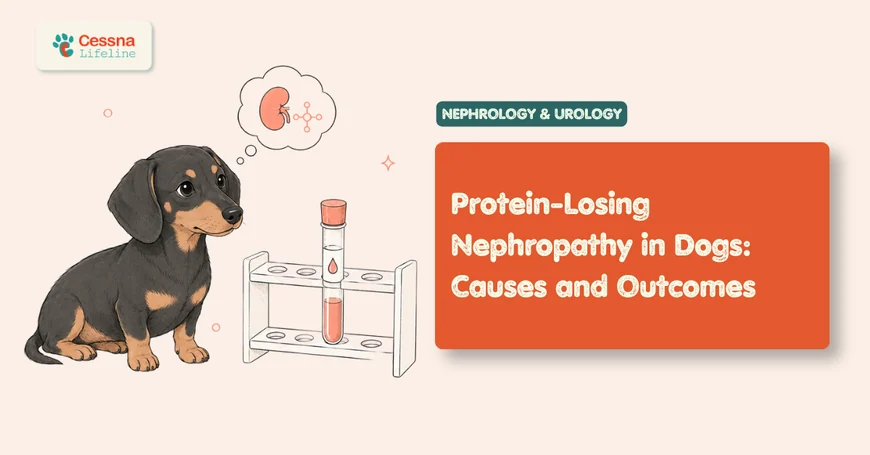
It comes up during a routine wellness visit. The bloodwork is normal. The dog looks healthy, eats well, has no obvious complaints. But the urinalysis, run almost as an afterthought, shows protein in the urine — more than there should be. The vet asks for a urine protein-to-creatinine ratio to confirm the finding.
That number changes everything. Not because the dog is visibly sick, but because proteinuria at that level is often the earliest detectable sign of a disease that, in its advanced form, causes fluid accumulation in the abdomen, life-threatening blood clots, and progressive kidney failure. The dog isn't in distress yet. It's the "yet" that matters.
Protein-losing nephropathy is one of the most consequential and underappreciated kidney diseases in veterinary medicine, not because it's rare, but because it announces itself so quietly. Understanding what it is, why it develops, and what the numbers on a lab report actually mean is the difference between catching it early and responding to a crisis.
What to Know Protein-losing nephropathy causes irreversible glomerular damage that progresses silently before producing clinical signs. A urine protein-to-creatinine (UPC) ratio above 2.0 in a dog is a threshold that demands urgent diagnostic workup. Without treatment, dogs with marked glomerular proteinuria have median survival times measured in months (ACVIM Consensus Guidelines on PLN, JVIM 2013). Caught early, progression can be slowed substantially.
What the Kidneys Are Losing — and Why It Matters
The glomerulus is the kidney's filtration unit, a coiled capillary bed surrounded by a specialized membrane designed to let water, electrolytes, and metabolic waste through while holding proteins back. Albumin, the blood's dominant protein, is large enough and negatively charged enough that a healthy glomerular membrane actively repels it. What ends up in the urine is a trace amount of small proteins, clinically negligible, that the tubules largely reclaim anyway.
What happens when that membrane fails? Everything downstream changes.
In protein-losing nephropathy, that membrane breaks down. The exact mechanism depends on the underlying disease — immune complexes lodging in the glomerular basement membrane, genetic defects in the collagen scaffolding, or amyloid deposits deforming the architecture — but the end result is the same. Albumin and other plasma proteins leak through into the filtrate and appear in the urine in quantities no healthy kidney would permit. The tubules can't reclaim it at scale. The protein is lost.
Why does losing protein in urine eventually threaten the whole dog? Because albumin does far more than occupy space in the bloodstream. It maintains oncotic pressure, the force that keeps fluid inside blood vessels rather than leaking into tissues. As serum albumin falls, that pressure drops. Fluid begins shifting out of the vascular compartment into the interstitium, the body cavities, and the limbs. Ascites, pleural effusion, and subcutaneous edema are the visible consequences of what started as an invisible number on a urinalysis.
At the same time, the liver, sensing the protein loss, accelerates synthesis of all its proteins — including clotting factors and lipoproteins. But it loses antithrombin III in the urine along with albumin, since antithrombin is a similar-sized molecule. Antithrombin keeps clotting in check. Without it, the dog enters a hypercoagulable state. Spontaneous thromboembolism becomes a genuine risk. Pulmonary thromboembolism is a recognized cause of acute respiratory distress and sudden death in dogs with advanced PLN, often occurring without warning.
Dogs with protein-losing nephropathy lose antithrombin III into the urine at rates paralleling albumin loss, creating a hypercoagulable state that significantly increases the risk of spontaneous thromboembolism. The ACVIM Consensus Guidelines on PLN recommend initiating antiplatelet therapy with clopidogrel when serum albumin falls below 2.0 g/dL to reduce this risk (Littman MP et al., JVIM 2013). Pulmonary thromboembolism is a documented cause of sudden deterioration and death in PLN dogs.
understanding dog bloodwork albumin levels
The Causes Behind PLN: From Immune Complexes to Breed Genetics
What causes the glomerular membrane to fail in the first place? The answer varies substantially, and it matters clinically because different underlying causes respond to different treatments — and some respond to none at all.
Immune-complex glomerulonephritis is the most common category, accounting for roughly half of PLN cases. The immune system deposits antigen-antibody complexes in the glomerular basement membrane, where they trigger complement activation and inflammatory damage. In many cases, the stimulus driving that immune activation is never identified — these are classified as idiopathic. In others, the trigger is a chronic disease elsewhere in the body: cancer, chronic infections, or immune-mediated conditions can all fuel the glomerular immune complex deposition that damages the filter.
What, then, triggers immune complex deposition in the first place? Sometimes it's an identifiable disease driving chronic immune activation. Often it isn't.
Infectious disease is a particularly important category among PLN causes, especially in geographically endemic areas. In North America, Lyme nephritis deserves special mention. Caused by Borrelia burgdorferi, the same organism responsible for Lyme disease, Lyme nephritis is a severe, rapidly progressive glomerulonephropathy that can also involve protein-losing enteropathy. Certain breeds are disproportionately affected, particularly Labrador Retrievers and Golden Retrievers, possibly due to immune response variation. Other tick-borne pathogens — Ehrlichia canis, Anaplasma species, and Rocky Mountain spotted fever — can also trigger glomerular inflammation through immune complex mechanisms. In Mediterranean and South American regions, canine leishmaniosis is a leading cause of immune-complex GN.
The Lyme nephritis paradox: Unlike most manifestations of Lyme disease, Lyme nephritis does not consistently resolve with antibiotic treatment alone. The glomerular immune complex damage can continue or progress even after Borrelia organisms are cleared. Dogs presenting with marked proteinuria and Lyme seropositivity require both doxycycline and aggressive management of the nephropathy itself — the antibiotic is necessary but not sufficient.
Familial and hereditary nephropathies form a distinct category driven not by infection or immune activation but by structural defects in the glomerular basement membrane, typically mutations affecting type IV collagen, the scaffold protein the membrane is built from. These conditions are breed-specific and often affect young dogs, sometimes progressing to end-stage kidney disease before age three.
Amyloidosis — the deposition of insoluble amyloid protein fibrils in the glomerular mesangium and eventually throughout the kidney — represents a separate mechanism entirely. It tends to produce some of the highest UPC ratios seen in clinical practice, sometimes exceeding 10 or 20, and carries the most guarded prognosis of any PLN category.
Immune-complex glomerulonephritis accounts for approximately 50% of PLN cases in dogs; familial/hereditary nephropathy for 15-20%; amyloidosis for 8-12%; and infectious disease-associated GN for 10-15%. Definitive diagnosis of the underlying mechanism requires renal biopsy with light microscopy, electron microscopy, and immunofluorescence (Littman MP et al., JVIM 2013; Vaden SL, JVIM 2011). Without histopathology, treatment decisions rest on probability rather than confirmed diagnosis.
<figcaption>PLN cause distribution in dogs. Immune-complex glomerulonephritis accounts for roughly half of all cases. Amyloidosis, while comprising only 10% of cases, carries the most guarded prognosis of any category. Sources: ACVIM Guidelines JVIM 2013; Vaden SL JVIM 2011.</figcaption> </figure>
Lyme disease nephritis in dogs
How PLN Is Detected: The UPC Ratio and What It Reveals
Proteinuria is not binary. All dogs have some protein in their urine; the question is how much, and whether the amount indicates glomerular leak. A standard urinalysis with a dipstick can flag protein qualitatively, ranging from a trace to 4+, but dipstick protein readings are unreliable for quantification and prone to false positives, particularly in concentrated urine. They're a screening signal, not a diagnosis.
The urine protein-to-creatinine ratio, or UPC, is the standard quantitative measure. Creatinine appears in urine at a concentration that reflects the urine's degree of dilution, so dividing urinary protein by urinary creatinine corrects for concentration and gives a number that's comparable across different urine samples regardless of how dilute or concentrated they are. A UPC below 0.5 is normal in dogs. Between 0.5 and 1.0 is borderline. Above 2.0, glomerular disease becomes the leading explanation.
Does the UPC ratio tell the whole story? Not alone. A single elevated UPC in a dog with active urinary tract inflammation, blood contamination from an in-heat female, or a traumatic catheter collection will be falsely elevated. IRIS guidelines recommend confirming persistent proteinuria with at least two UPC measurements from first-morning samples collected two to four weeks apart, from a dog with a clean collection and no concurrent lower urinary tract inflammation, before concluding the elevation reflects glomerular disease.
<figcaption>UPC ratio severity scale in dogs. IRIS defines proteinuria substage P3 at UPC above 2.0. Dogs with amyloidosis frequently present with UPC above 10, sometimes exceeding 20-30. Sources: IRIS Kidney 2023; ACVIM PLN Guidelines 2013.</figcaption> </figure>
Why not simply treat a high UPC without investigating further? Because PLN has multiple causes that require completely different treatments, and treating the wrong mechanism wastes time the kidneys don't have.
Once a persistently elevated UPC is confirmed, the next step is characterizing which part of the urinary tract the protein is coming from. Lower urinary tract disease, such as cystitis or urethral inflammation, can cause mild to moderate proteinuria. True glomerular proteinuria tends to be more marked and is dominated by albumin on urine protein electrophoresis, because albumin is the protein the glomerular barrier normally retains most strictly. When the UPC exceeds 2.0 with urinary sediment showing only occasional red or white cells and no evidence of lower tract disease, a renal source is essentially confirmed.
urine protein-to-creatinine ratio in dogs
The Complications That Follow Protein Loss
Understanding the UPC ratio is only half the picture. What happens inside the dog as the protein keeps leaving is what determines the urgency of the situation.
Hypoalbuminemia is the most direct downstream consequence. The liver synthesizes albumin continuously but cannot keep pace with losses driven by severe glomerular disease. As the serum albumin falls, the oncotic pressure holding fluid in the vascular space declines. At what point does this become clinically visible? Generally below 2.0 g/dL, fluid begins accumulating in dependent spaces. Below 1.5 g/dL, ascites, pleural effusion, and limb edema become likely. These are not the primary danger — they're the visible marker that the disease has been advancing silently for some time.
The more immediately dangerous complication is thromboembolism. Loss of antithrombin III mirrors albumin loss, because the two molecules have similar sizes and are filtered and lost together. Antithrombin is the body's primary brake on the coagulation cascade. Without it, clot formation becomes pathologically favored. Pulmonary thromboembolism is the most feared consequence — a blood clot that lodges in the pulmonary vasculature, causing acute respiratory distress that can look identical to pneumonia or heart failure and is frequently fatal before diagnosis is confirmed.
<figcaption>Serum albumin thresholds and corresponding clinical actions in dogs with PLN. Antiplatelet therapy is recommended at or below 2.0 g/dL. Below 1.5 g/dL, clinical decompensation is likely and hospitalisation should be considered. Source: ACVIM PLN Guidelines 2013.</figcaption> </figure>
The ACVIM Consensus Guidelines on PLN specify serum albumin below 2.0 g/dL as the threshold for initiating clopidogrel antiplatelet therapy, based on the correlation between antithrombin III loss (co-excreted with albumin) and thromboembolic risk. Below 1.5 g/dL, pleural effusion, ascites, and spontaneous thromboembolism become highly probable clinical complications (Littman MP et al., JVIM 2013).
thromboembolism risk in dogs with kidney disease
Breed Predispositions: Dogs Born at Higher Risk
For some dogs, PLN isn't triggered by infection or immune dysregulation. It's written into the genome. Several breeds carry inherited defects in the structural proteins of the glomerular basement membrane, and these dogs often develop nephropathy young — in some cases, before their third birthday.
The Soft-Coated Wheaten Terrier has one of the most extensively documented hereditary nephropathies. Affected dogs develop a dual syndrome: protein-losing nephropathy and protein-losing enteropathy simultaneously, losing protein from both the kidneys and the gastrointestinal tract. This double burden accelerates hypoalbuminemia beyond what either condition alone would produce. The genetic basis has been associated with mutations in podocin and other slit diaphragm proteins critical to glomerular filtration integrity. Breeding programs within the breed have improved but not eliminated the prevalence of affected dogs.
The Samoyed carries an X-linked hereditary nephritis that parallels human Alport syndrome: a mutation in the gene encoding type IV collagen, the structural protein that forms the scaffold of the glomerular basement membrane. Affected male Samoyeds (who have only one X chromosome) progress to renal failure early, often by 18 months to 3 years, while carrier females may show only persistent proteinuria for years before developing azotemia. Bull Terriers carry a similar collagen IV defect, inherited autosomal-recessively, producing what is termed Bull Terrier hereditary nephritis.
Which breeds are most vulnerable? Beyond these three, English Cocker Spaniels, Bernese Mountain Dogs (where hereditary glomerulonephritis is well-documented and rapidly progressive), and Rottweilers all appear in the hereditary nephropathy literature with varying degrees of penetrance and severity. Among breeds not classically predisposed, Labrador Retrievers and Golden Retrievers are disproportionately represented in Lyme nephritis case series, likely due to immune response characteristics rather than structural kidney defects.
Genetic testing is now available for several of these breeds. The Soft-Coated Wheaten Terrier Club of America has supported a prospective health screening program, and DNA tests for podocin mutations exist. For buyers of Samoyed, Bull Terrier, or English Cocker Spaniel puppies, asking whether parents were tested for hereditary nephropathy is not overcaution — it's the only way to know whether a puppy is starting life with a glomerular basement membrane that will hold.
Soft-Coated Wheaten Terriers can develop simultaneous protein-losing nephropathy and protein-losing enteropathy through defects in podocin and related slit-diaphragm proteins; Samoyeds and Bull Terriers carry X-linked or autosomal forms of hereditary nephritis analogous to human Alport syndrome through mutations in type IV collagen genes; Bernese Mountain Dogs develop rapidly progressive hereditary glomerulonephritis (Vaden SL, JVIM 2011; ACVIM PLN Guidelines, JVIM 2013).
hereditary kidney disease in dog breeds
Treatment and Monitoring: Slowing the Progression
How does anyone know which treatment to use without a biopsy? Strictly speaking, they don't. The ACVIM Consensus Guidelines are explicit: definitive treatment of immune-complex glomerulonephritis requires histopathologic confirmation, because different patterns — membranous nephropathy, membranoproliferative GN, focal segmental glomerulosclerosis — respond differently to immunosuppressive protocols. Treating all PLN with the same immunosuppressive regimen is both ineffective and potentially harmful. Renal biopsy with light microscopy, immunofluorescence, and electron microscopy is the only way to distinguish these patterns.
That said, several elements of PLN management apply broadly, regardless of the biopsy diagnosis.
ACE inhibitors and angiotensin receptor blockers reduce intraglomerular pressure and decrease the amount of protein leaking through the damaged filter. Enalapril and benazepril are the most commonly used ACE inhibitors in dogs with PLN; telmisartan, an angiotensin receptor blocker, has shown particular promise for proteinuria reduction and is increasingly included in treatment protocols. These drugs do not repair the glomerulus. They reduce the filtration pressure driving protein loss, buying time for other interventions and slowing progression. A 30-50% reduction in UPC ratio is a reasonable response target over two to three months.
What happens when anti-proteinuric therapy isn't enough to hold the UPC stable? That's when the biopsy result determines whether immunosuppression is appropriate and which protocol to follow.
Omega-3 fatty acids (EPA and DHA from fish oil) have a modest but real anti-proteinuric effect and anti-inflammatory benefit, and are included in ACVIM recommendations for all PLN patients at doses of approximately 0.2-0.3 g EPA+DHA per kg of body weight daily. Dietary protein management requires careful balance: severely azotemic PLN dogs may benefit from protein restriction to reduce uremic load, while hypoalbuminemic dogs need adequate protein to support albumin synthesis. This is one of the most contested aspects of PLN nutrition management, and the answer frequently depends on which problem is more severe.
From the consultation room: Owners often ask whether there's something the dog ate that caused this, or something they can change about the environment. For hereditary forms and idiopathic immune-complex GN, the honest answer is no — nothing in the diet or environment caused it and nothing non-medical will stop it. What owners can control is the consistency of monitoring: a UPC rechecked every four weeks during active management is the signal that lets clinicians know whether the treatment is working before another clinical crisis develops.
For dogs with immune-complex GN confirmed on biopsy, immunosuppressive therapy is added. Mycophenolate mofetil and chlorambucil are the agents most commonly used in current protocols, largely replacing azathioprine based on more favorable efficacy and side effect profiles. The treatment decision also depends on the biopsy pattern: membranous GN may respond well to immunosuppression; focal segmental glomerulosclerosis responds less predictably. Dogs with hereditary nephropathy and amyloidosis generally do not respond to immunosuppression and are managed with anti-proteinuric and supportive measures.
Telmisartan, an angiotensin receptor blocker, demonstrated a significant reduction in proteinuria in dogs with PLN in clinical trials and is now included in ACVIM treatment recommendations alongside ACE inhibitors. Omega-3 fatty acids at 0.2-0.3 g EPA+DHA per kg daily and antiplatelet therapy with clopidogrel (when albumin < 2.0 g/dL) are recommended for all PLN patients (ACVIM Consensus Guidelines on PLN, JVIM 2013; Brown SA et al.).
ACE inhibitors and ARBs for kidney disease in dogs
Frequently Asked Questions
Is protein in a dog's urine always a sign of kidney disease?
Not always. Mild transient proteinuria can follow intense exercise, fever, stress, or urinary tract inflammation. A single elevated protein reading on a dipstick is a flag, not a diagnosis. Persistent proteinuria confirmed on two separate UPC measurements from clean samples, at least two to four weeks apart, with no concurrent lower urinary tract disease, is what raises the index of suspicion for glomerular kidney disease. Dipstick proteinuria alone, particularly in concentrated urine, is frequently a false positive.
What does a UPC ratio of 3.0 mean for my dog?
A UPC of 3.0 places the dog solidly in the IRIS proteinuria substage P3, indicating significant glomerular protein loss well above the normal threshold of 0.5. It warrants a complete workup including bloodwork, blood pressure measurement, abdominal ultrasound, urine culture, and infectious disease serology, and should be confirmed with a second measurement before initiating treatment. It does not by itself indicate how long the disease has been present, what caused it, or how fast it will progress — those questions require further diagnostics.
Do all dogs with PLN need a kidney biopsy?
Not always, but it's strongly recommended when immunosuppressive therapy is being considered. Biopsy is the only way to distinguish immune-complex GN subtypes, hereditary nephropathy, and amyloidosis — three conditions that overlap in clinical presentation but diverge sharply in treatment. Without biopsy, immunosuppression is started based on probability rather than confirmed diagnosis, and in a dog with amyloidosis or hereditary nephropathy, immunosuppression adds toxicity without benefit. The risk of the biopsy procedure must be weighed against the platelet count, albumin level, blood pressure, and degree of azotemia.
Can PLN be cured?
Cure is possible when PLN is driven by a treatable underlying cause. A dog with early Lyme nephritis treated promptly with doxycycline and ACE inhibitors may normalize UPC and maintain kidney function. A dog with immune-complex GN secondary to a resolved neoplasm may show complete remission after cancer treatment. For idiopathic immune-complex GN, amyloidosis, and hereditary nephropathy, the goal is slowing progression and managing complications rather than cure. These are diseases managed over months to years, not resolved in weeks.
How long do dogs with PLN live?
Prognosis varies widely based on UPC at diagnosis, presence of azotemia, underlying cause, and response to treatment. Dogs with non-azotemic PLN and responsive immune-complex GN can have survival times measured in years with appropriate management. Dogs presenting with concurrent azotemia and marked hypoalbuminemia have significantly shorter median survival times. Amyloidosis carries the most guarded prognosis, with most dogs surviving fewer than 12 months after diagnosis. Regular monitoring allows early detection of deterioration, which is why UPC and albumin rechecks every four to eight weeks during active management are so important.
Before the Swelling Begins
The most powerful thing a UPC ratio offers is time. By the time ascites is visible, by the time a dog's legs are edematous, by the time a dog collapses with a pulmonary embolism, the glomerular disease driving these events has usually been measurable — and potentially treatable — for months.
The UPC ratio is not a scary number. It's an early warning. A dog with a UPC of 2.5 and normal albumin and creatinine has a window that a dog in hypoalbuminemic decompensation has already closed. That window is where treatment works best: where immunosuppression, if the biopsy supports it, can slow the immune complex deposition; where telmisartan and fish oil can reduce the protein loss that will eventually deplete the albumin; where antiplatelet therapy, started before the albumin falls below 2.0, prevents the thromboembolism that arrives without warning.
Protein-losing nephropathy is not a disease that announces itself urgently. It takes its time. So should the response — measured, specific, based on what the biopsy shows and what the numbers are doing, with rechecks frequent enough to catch the trajectory before it becomes irreversible.
The swelling, when it comes, means the window has narrowed. The UPC ratio, run during a routine wellness visit with no symptoms in sight, is the test that catches the disease before that happens.