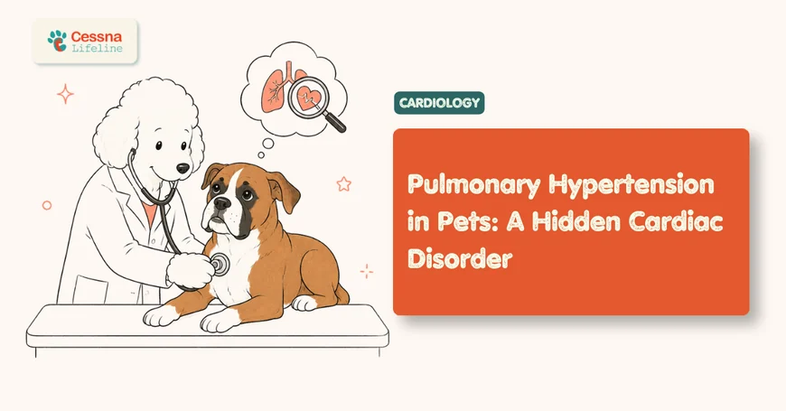
Pulmonary Hypertension in Pets: A Hidden Cardiac Disorder
Pulmonary hypertension affects 34% of dogs referred for cardiac evaluation. Recognize exercise intolerance, respiratory distress, and breathlessness causes.
Your dog slows on the stairs. She stops midway through a walk she used to finish without a second thought. You chalk it up to aging or summer heat. Then one morning she collapses in the yard.
Pulmonary hypertension (PH) is a condition in which blood pressure inside the pulmonary arteries, the vessels carrying blood from the right ventricle into the lungs, climbs above normal thresholds. The right side of the heart compensates by working harder until it can't. What makes PH particularly dangerous is how quietly it builds: the early signs are vague, easily dismissed, and easy to attribute to a dozen other things.
Understanding what PH is, what causes breathlessness in dogs, and how exercise intolerance and respiratory distress appear in practice gives owners a genuine chance to catch this condition before it reaches a breaking point.
Key Takeaways
- Pulmonary hypertension was diagnosed in 34.3% of dogs referred for echocardiographic cardiac evaluation (ScienceDirect, 2019).
- Exercise intolerance is the most commonly reported early sign and often appears months before overt respiratory distress.
- Treated dogs reach a median survival of approximately 567 days, with some living beyond 5 years (JVIM, 2020).
- Echocardiography by a veterinary cardiologist is the only reliable way to confirm PH.
Table of Contents
- What Is Pulmonary Hypertension in Pets?
- Why Does Pulmonary Hypertension Go Undetected?
- What Causes Breathlessness in Dogs?
- Cardiac Causes
- Respiratory Causes
- Other Systemic Causes
- How Does Exercise Intolerance Signal Pulmonary Hypertension?
- What Are the Signs of Respiratory Distress in Pets?
- How Is Pulmonary Hypertension Diagnosed in Pets?
- What Treatment Options Exist for Pets with PH?
- Frequently Asked Questions
- Conclusion: Acting Before the Crisis
What Is Pulmonary Hypertension in Pets?
Pulmonary hypertension is defined by a sustained elevation in pulmonary arterial pressure above 25 mmHg at rest in dogs, a threshold at which the right ventricle begins to remodel under the increased afterload (ACVIM Consensus Statement, JVIM 2020). PH isn't a disease in itself but a hemodynamic consequence of something else failing upstream, whether in the heart, the lungs, or the systemic circulation.
The ACVIM classifies canine PH into five mechanistic groups:
- Group 1 (Pulmonary Arterial Hypertension, PAH): Intrinsic disease of the pulmonary vessel walls; rare in dogs and cats but carries the poorest prognosis.
- Group 2 (Post-capillary PH): Pressure backs up from left-sided heart disease into the pulmonary veins and onward. This is the most common form in small-breed dogs.
- Group 3 (Hypoxic PH): Chronic low oxygen from respiratory disease drives compensatory vasoconstriction in the lungs, thickening vessel walls over time.
- Group 4 (Thromboembolic PH): Clots physically obstruct pulmonary arteries and spike resistance acutely.
- Group 5 (Unclear/multifactorial): Mixed mechanisms or rare systemic conditions like vasculitis.
<figcaption>PH prevalence varies by population: from 34.3% in general cardiac cohorts to 60% in West Highland White Terriers with pulmonary fibrosis.</figcaption> </figure>
Cats develop PH too, most commonly secondary to hypertrophic cardiomyopathy or chronic lower airway disease. They present with the same broad syndrome but tend to mask signs even more effectively than dogs, making early detection harder.
The ACVIM Consensus Statement recommends Doppler echocardiography as the primary non-invasive screening tool for PH in dogs, with a tricuspid regurgitation (TR) velocity above 2.8 m/s used as the threshold for mild-to-moderate disease (Reinero et al., JVIM 2020). Groups 2 and 3, driven by left heart disease and respiratory hypoxia, account for the vast majority of cases seen in clinical practice.
degenerative mitral valve disease in dogs
Why Does Pulmonary Hypertension Go Undetected?
PH slips past owners for one straightforward reason: its early signs look exactly like normal aging. A dog that was once fast and now trots, that coughs once at night, that prefers lying still over chasing a ball; none of those changes sound like a cardiovascular emergency. But collectively, they represent a right ventricle under growing pressure.
The disease progresses in phases. Early on, the right ventricle compensates by contracting harder. Resting breathing rate creeps up by a few breaths per minute. The dog may cough occasionally, especially at night or after lying down. She's still functional. She might even appear fine on a routine physical exam because a stethoscope cannot detect elevated pulmonary arterial pressure in most cases.
By the time obvious signs appear, including fainting during exercise, a visibly distended abdomen, blue-tinged gums, or persistent labored breathing, the right ventricle is already under severe chronic strain.
What the data tells us: 60% of West Highland White Terriers with canine idiopathic pulmonary fibrosis had moderate-to-severe PH at the time of evaluation (BMC Veterinary Research, 2021), yet the majority of those dogs had been managed by general practitioners for months under the assumption that slow respiratory worsening was simply disease progression, not secondary PH layered on top.
The practical consequence: owners and vets who don't actively look for PH tend to find it late, when the treatment window has narrowed.
monitoring dogs with chronic heart disease at home
What Causes Breathlessness in Dogs?
Breathlessness in dogs (dyspnea) has a wide differential, and cardiac disease is not always the top of the list. A retrospective study of 229 dogs referred for dyspnea evaluation found that upper airway disease (32%) and lower respiratory tract disease (33%) were the most common causes, with cardiac disease accounting for only 12% of cases (JAVMA, 2011).
<figcaption>Source: JAVMA 2011 retrospective study of 229 dogs referred for dyspnea evaluation</figcaption> </figure>
This distribution matters because it shapes the diagnostic approach. Before anyone concludes that a breathless dog has heart disease, the airways and pleural space must be assessed. That said, when breathlessness does stem from cardiac or pulmonary vascular disease, PH is frequently the underlying mechanism.
Cardiac Causes
Left-sided heart disease is the most common cardiac trigger for breathlessness in dogs. When the mitral valve degenerates, blood backs up into the pulmonary veins. Pressure rises in the pulmonary capillaries, fluid leaks into the alveoli (cardiogenic pulmonary edema), and the dog breathes faster to compensate. Cavalier King Charles Spaniels, Dachshunds, Maltese, and other small breeds carry genetic predisposition to degenerative mitral valve disease, the same condition responsible for Group 2 PH.
Dilated cardiomyopathy (DCM), which preferentially affects large breeds like Doberman Pinschers, Irish Wolfhounds, and Great Danes, causes breathlessness through a different pathway: a weakened left ventricle fails to pump adequate forward output, raising back-pressure through the entire pulmonary circuit.
Respiratory Causes
Chronic bronchitis, tracheal collapse, brachycephalic airway syndrome, interstitial lung disease, and pulmonary fibrosis all restrict airflow and reduce blood oxygen levels. The resulting hypoxia triggers vasoconstriction of the pulmonary arterioles as a compensatory mechanism that, paradoxically, worsens pulmonary pressure over time. Heartworm disease (Dirofilaria immitis) deserves specific mention: adult worms physically reside in the pulmonary arteries, provoking both mechanical obstruction and vascular inflammation. Dogs successfully treated for heartworm can still carry residual PH from permanent vessel remodeling.
Other Systemic Causes
Pulmonary thromboembolism can cause sudden, severe respiratory distress in dogs that appeared stable hours earlier. High-risk populations include dogs with protein-losing nephropathy, hyperadrenocorticism (Cushing's disease), or immune-mediated hemolytic anemia. Anemia itself reduces oxygen-carrying capacity and triggers compensatory hyperpnea. Obesity compounds any pre-existing airway or cardiac issue by adding mechanical load to every breath.
Upper airway and lower respiratory tract diseases together account for 65% of breathlessness cases in dogs examined for dyspnea, while cardiac disease drives approximately 12% (JAVMA, 2011). This distribution means that treating breathlessness as a cardiac problem by default, without thoracic radiography and airway assessment, risks missing the correct diagnosis in the majority of cases.
How Does Exercise Intolerance Signal Pulmonary Hypertension?
Exercise intolerance is the most commonly reported early sign of PH in dogs and the one most often dismissed as normal aging or mild obesity. When pulmonary arterial pressure is elevated, the right ventricle cannot increase its output during exertion the way a healthy heart can. Blood delivery to working muscles is blunted, oxygen debt accumulates faster, and the dog stops, sits, or simply refuses to continue activity it used to handle with ease.
So what's the difference between a dog that's slowing with age and one with true exercise intolerance? Context and proportion. An older healthy dog takes more time but still completes familiar routes. A dog with PH stops at unpredictable, early points in the walk, shows disproportionate panting for minimal effort, and often takes longer than expected to return to normal breathing afterward.
Clinical signs documented in dogs with respiratory-associated PH treated with sildenafil include exercise intolerance as a primary presenting complaint, followed by coughing, tachypnea, and in more advanced cases, exertional syncope (Johnson et al., JVIM 2020). Exertional syncope, where the dog faints during or immediately after activity, is the most alarming presentation. The mechanism is direct: during exertion, cardiac output demand rises sharply. With fixed pulmonary vascular obstruction, the right ventricle can't meet that demand. Systemic blood pressure drops briefly, cerebral perfusion falls, and the dog loses consciousness.
From clinical practice: The owners who catch PH earliest in our experience are those who notice something specific, not just "she's slower." The tell is a dog that stops at the same point every walk, sits down, and looks uncomfortable for 30 to 60 seconds before she's willing to move again. That pattern of predictable early exercise cessation is different from normal tiredness.
Syncope during exercise should be treated as a cardiac emergency regardless of the suspected cause. Any dog that collapses after physical activity needs urgent evaluation, not a "wait and see" approach.
Exertional syncope and labored respiration following activity are clinical signs frequently directly attributable to pulmonary hypertension, according to the ACVIM Consensus Statement (Reinero et al., JVIM 2020). Dogs that present with exercise-induced collapse typically have more advanced right ventricular dysfunction at echocardiography than those presenting for exercise intolerance alone, reinforcing the importance of referral before syncope occurs.
episodic weakness and syncope in dogs
What Are the Signs of Respiratory Distress in Pets?
Respiratory distress in pets exists on a spectrum, from increased resting respiratory rate at the mild end to cyanosis and collapse at the severe end. PH tends to announce itself through the subtle end first, which is precisely why owners miss it.
A healthy resting dog breathes 15 to 30 times per minute. Most veterinary cardiologists ask owners of at-risk dogs to count resting breaths once per week and log them. A consistent rate above 30 breaths per minute, or a trend of rising numbers over weeks, should prompt a vet call the same day rather than waiting for the next scheduled appointment.
Signs that respiratory distress has progressed to a level requiring urgent same-day evaluation include:
- Abdominal distension (ascites): Fluid accumulating from right heart failure; the belly looks round and taut, and the dog may be reluctant to lie flat.
- Cyanosis: Blue or purplish gums indicate that oxygen saturation has fallen to dangerous levels. This is a genuine emergency.
- Orthopnea: The pet refuses to lie down and rests in a sternal or sitting position because lying flat worsens breathing mechanics.
- Abdominal effort during breathing: The dog uses belly muscles to push air out, which signals that respiratory muscles are fatiguing.
- Extended neck posture: Stretching the neck forward during breathing helps open the airway but reflects significant respiratory effort.
Cats are particularly challenging here. Respiratory distress signs in cats with PH can be as subtle as hiding more, eating less, or breathing with a slightly elevated effort after being picked up. Any open-mouth breathing in a resting cat should be treated as an emergency without waiting to see if it resolves on its own.
What owners miss most: Orthopnea in dogs is underrecognized because owners don't notice that their pet is always sitting up. Once it's pointed out, many say the dog hasn't voluntarily lain on its side in weeks. That observation alone warrants an urgent echocardiographic referral.
Respiratory distress in PH progresses from mild tachypnea to overt cyanosis as right ventricular function deteriorates (ACVIM Consensus Statement, JVIM 2020). Dogs with cyanotic mucous membranes at presentation have significantly higher pulmonary pressures and shorter survival times than those identified at the exercise-intolerant stage, making the resting respiratory rate the most practical early warning tool available to owners.
home monitoring for dogs with heart disease
How Is Pulmonary Hypertension Diagnosed in Pets?
The gold standard for diagnosing PH in dogs is Doppler echocardiography, which almost always means a referral to a veterinary cardiologist. A standard physical examination can raise suspicion, finding an unusual split S2 heart sound, rough lung sounds, jugular venous distension, or a cardiac murmur, but it can't measure pulmonary arterial pressure.
The echocardiogram measures the velocity of the tricuspid regurgitation (TR) jet. A TR velocity above 2.8 m/s correlates with elevated pulmonary pressure and indicates at least mild-to-moderate PH. Values above 3.4 m/s signal severe disease. The right ventricle dimensions, the degree of right atrial enlargement, and whether the interventricular septum has flattened (indicating pressure equalization between chambers) all add additional detail about disease severity.
A complete diagnostic workup typically includes:
- Thoracic radiographs: Detect pulmonary infiltrates, cardiomegaly, enlarged main pulmonary artery segment, or pleural effusion.
- Complete blood count and chemistry: Screen for concurrent systemic disease driving PH.
- Heartworm antigen test: Mandatory in endemic regions for any dog with unexplained PH.
- NT-proBNP blood test: A biomarker that rises with cardiac wall stress; an elevated level increases clinical suspicion and helps triage referral urgency.
- Doppler echocardiography: Definitive. Measures TR velocity, right and left ventricular function, and identifies structural cardiac disease.
- CT pulmonary angiography: Used when thromboembolic PH is suspected and clot burden needs to be mapped before anticoagulation decisions.
A 42.1% PH prevalence among dogs with DMVD Stage C heart disease underscores how often PH coexists with known cardiac conditions and goes undetected without dedicated echocardiographic screening (ResearchGate, 2020). Every dog in Stage C or with unexplained breathlessness and exercise intolerance warrants specialist-level cardiac imaging.
veterinary cardiology referral guide
What Treatment Options Exist for Pets with PH?
Treatment for PH in dogs focuses on two parallel goals: reducing pulmonary vascular resistance and addressing the underlying cause. Sildenafil, a phosphodiesterase-5 (PDE5) inhibitor originally developed for human pulmonary arterial hypertension, is the most widely used PH-specific drug in veterinary medicine. It dilates the pulmonary vasculature, lowers pulmonary arterial pressure, and reduces right ventricular afterload.
For dogs with left-heart-driven PH (Group 2), the approach pairs standard cardiac medications with PH-specific therapy. Pimobendan improves left ventricular contractility and reduces back-pressure. ACE inhibitors reduce volume load. Sildenafil then targets the pulmonary arterial pressure elevation directly. A 2022 retrospective study found that dogs with DMVD-associated PH who received sildenafil had significantly longer survival times than those on standard cardiac medications alone (Taylor and Francis, 2022).
<figcaption>Estimated survival trajectory for dogs with treated pulmonary hypertension: 84% at 6 months, 73% at 12 months, with median survival approximately 567 days.</figcaption> </figure>
Current evidence shows that treated dogs reach 84% survival at 6 months and 73% survival at 12 months when stable in the first week of therapy (AskAVet, 2025). The median overall survival is approximately 567 days from diagnosis. Some dogs in published respiratory-associated PH cohorts were alive 5 years after diagnosis (JVIM, 2020).
Additional treatment components include:
- Oxygen supplementation: At-home oxygen concentrators reduce hypoxic vasoconstriction in dogs with respiratory-driven PH and can meaningfully improve comfort.
- Diuretics (furosemide, spironolactone): Manage fluid accumulation from right heart failure and reduce work of breathing.
- Activity restriction: Strenuous exercise, stairs, and off-leash running should be limited to prevent exertional syncope.
- Anticoagulation: For thromboembolic PH, clopidogrel or low-molecular-weight heparin addresses the clot burden.
- Follow-up echocardiography: Monthly until the dog is stable, then every 3 to 6 months to track right ventricular dimensions and TR velocity trends.
Treatment also depends heavily on resolving the primary cause. A dog with heartworm-induced PH needs heartworm treatment alongside sildenafil. A dog with DMVD needs optimal left-heart management. Addressing only the PH and ignoring the driver rarely produces durable results.
Dogs with respiratory-associated PH treated with sildenafil showed 50% mortality at 6 months post-study entry in a 25-dog cohort, yet 4 dogs were alive 5 years after diagnosis (Johnson et al., JVIM 2020). Early response to sildenafil in the first 4 weeks and the nature of the underlying cause are the strongest predictors of long-term survival in published veterinary literature.
cardiac medications for dogs
Frequently Asked Questions
Can cats develop pulmonary hypertension?
Yes. Cats develop PH most often secondary to hypertrophic cardiomyopathy or chronic lower airway disease. The diagnostic approach mirrors dogs: Doppler echocardiography is required for confirmation. Any open-mouth breathing in a resting cat constitutes an emergency, since cats with cardiac-origin respiratory distress can decompensate rapidly. Sildenafil has been used off-label in cats with anecdotal benefit, though controlled feline data remain limited.
How quickly does pulmonary hypertension progress in dogs?
Progression depends entirely on the underlying cause. PH from mitral valve disease builds over months to years as the heart disease advances through staging. Thromboembolic PH can develop within hours of a large clot event. Without treatment, dogs in severe PH rarely survive beyond weeks to a few months from the time of diagnosis. With sildenafil therapy, median survival in published cohorts reaches approximately 567 days (JVIM, 2020), though individual outcomes vary considerably.
Is any exercise safe for dogs with pulmonary hypertension?
Low-intensity, leash-controlled activity is generally acceptable. Short, calm walks on flat ground give dogs mental stimulation without triggering dangerous exertional stress. The hard restrictions are strenuous play, stair climbing, off-leash running, and anything that produces heavy panting. Dogs with moderate-to-severe PH should be rechecked by echocardiography before any increase in activity level is permitted, since a drop in TR velocity during follow-up is the most reliable sign that PH is responding to treatment.
Which dog breeds carry the highest risk for pulmonary hypertension?
Small-breed dogs prone to degenerative mitral valve disease carry the highest group-level risk, particularly Cavalier King Charles Spaniels, Maltese, and Dachshunds. West Highland White Terriers face elevated risk specifically from idiopathic pulmonary fibrosis, with 60% of affected individuals developing moderate-to-severe PH (BMC Veterinary Research, 2021). Large breeds with DCM, including Doberman Pinschers and Irish Wolfhounds, develop PH through left ventricular failure. Brachycephalic breeds with severe airway obstruction represent another distinct at-risk group.
Can pulmonary hypertension be cured in dogs?
A functional cure is possible only when the underlying cause is fully reversible, such as successful treatment of an acute thromboembolism in an otherwise healthy dog. In most clinical scenarios, PH is managed rather than cured. The goal of therapy is to maintain quality of life, slow right ventricular remodeling, and extend comfortable functional time. Many dogs achieve good quality of life for a year or more with consistent treatment and regular monitoring (JVIM, 2020).
quality of life assessment for dogs with chronic cardiac disease
Conclusion: Acting Before the Crisis
Pulmonary hypertension does not arrive with a single, unmistakable moment. It accumulates quietly behind exercise sessions your dog stops a little early, behind a breathing rate that ticks up three beats per minute month after month, behind a dog that just isn't quite herself.
The tools to catch it early are surprisingly accessible. A 15-second weekly resting respiratory rate count at home. A prompt vet call for any dog showing predictable exercise cessation or a persistent rate above 30 breaths per minute. An echocardiographic referral before syncope forces the issue.
Modern therapy gives many affected dogs a year or more of functional, comfortable life after diagnosis. That outcome depends on how early the right ventricle is identified as a system under strain. The earlier the catch, the wider the treatment window, and the longer the dog and her family have together.