Canine Atopic Dermatitis: Environmental Allergies in Dogs
Atopic dermatitis affects 10 to 15% of dogs and causes chronic itching with no fleas involved. Learn how vets diagnose and treat environmental allergies in dogs.
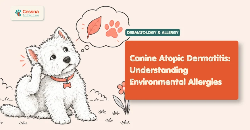
A dog that scratches its ears daily, chews its paws until the fur turns rust-colored from saliva staining, and rubs its face on the carpet is not a dog that needs a flea treatment. It is a dog with a probable diagnosis of atopic dermatitis: an inherited predisposition to develop immunological hypersensitivity to environmental allergens that, in most cases, are inhaled or absorbed through the skin rather than ingested. The itching is not behavioral, it is not nutritional in origin, and it does not resolve with topical antiparasitic treatment. It requires a dermatological diagnosis and a long-term management strategy. food allergy in dogs vs. environmental allergy guide
What to Know Atopic dermatitis is a genetic predisposition to become sensitized to environmental allergens including dust mites, pollens, and molds. It affects 10 to 15% of dogs, typically begins between 6 months and 3 years of age, and causes chronic recurrent pruritus centered on the face, paws, groin, and ears. It is manageable but rarely curable without long-term treatment commitment.
The Itch That Comes Back Every Year
The clinical history that alerts a veterinarian to atopic dermatitis is not just the presence of pruritus but its pattern. A dog that itches primarily in spring and summer, or whose symptoms worsen every August without a clear external trigger, is a dog whose itch is following an allergen calendar. Seasonal pollen counts rise and fall. House dust mite concentrations peak in autumn when heating systems recirculate household air. Mold spore counts correlate with humidity and leaf litter accumulation. These are not coincidences; they are the environmental fingerprint of atopy.
The condition typically manifests between 6 months and 3 years of age. A dog that develops significant pruritus for the first time at age 7 with no prior history is less likely to have atopy and more likely to have a newly acquired contact allergen, an internal disease with cutaneous manifestation, or a parasitic cause. Atopy in dogs is a diagnosis that almost always has its roots in early adulthood. The symptoms may worsen with age as sensitization broadens to include additional allergens, but the initial presentation is nearly always in the first three years of life.
Breed predisposition is well-established. West Highland White Terriers, Golden Retrievers, Labrador Retrievers, English Bulldogs, French Bulldogs, German Shepherds, Boxers, and Shih Tzus are disproportionately represented in atopy populations. Breed predisposition does not guarantee the diagnosis, but it raises the pre-test probability enough that atopy should be the leading differential in a pruritic Westie of any age.
What Causes Atopic Dermatitis in Dogs?
The pathophysiology of canine atopic dermatitis involves two concurrent defects: an intrinsic abnormality in the skin barrier that allows allergen penetration, and an immune dysregulation that shifts the response to those allergens toward a Th2-dominant hypersensitivity pattern. Neither defect alone produces clinical disease; the combination is necessary. dog skin infections pyoderma guide
Skin barrier dysfunction: The outermost layer of the skin (stratum corneum) in atopic dogs has reduced ceramide content, altered lipid composition, and increased transepidermal water loss compared to non-atopic dogs. This structural compromise allows allergen proteins to penetrate the epidermis at concentrations that exceed what an intact barrier would permit, sensitizing resident immune cells (Langerhans cells, dendritic cells) to allergen proteins they would not encounter in a healthy barrier.
Immune dysregulation: Once sensitized, subsequent allergen exposure triggers IgE-mediated mast cell degranulation and Th2 cytokine release (IL-4, IL-13, IL-31). IL-31 is the primary pruritogenic cytokine in canine atopy: it directly binds receptors on sensory neurons in the skin, generating the itch signal. This mechanistic detail is clinically significant because it is the target of lokivetmab (Cytopoint), a monoclonal antibody that neutralizes circulating IL-31 before it reaches its neuronal receptor.
Allergen sources: The most common environmental allergens identified by testing in atopic dogs include house dust mites (Dermatophagoides farinae and D. pteronyssinus), storage mites (Tyrophagus putrescentiae), grass pollens (Bermuda, Timothy, Kentucky Blue), tree pollens (oak, birch, ash), weed pollens (ragweed, mugwort), and mold spores (Aspergillus, Alternaria, Cladosporium). Most atopic dogs are sensitized to multiple allergen groups simultaneously, which is why symptoms often persist year-round even in dogs with initial seasonal presentations as sensitization broadens over time.
Citation Capsule: Canine atopic dermatitis affects an estimated 10 to 15% of the dog population and is the second most common skin condition seen in veterinary practice after flea allergy dermatitis; the condition has a recognized heritable component with multiple breed predispositions, and concordance studies in related dogs demonstrate genetic transmission of both the skin barrier abnormality and the immune dysregulation that together produce clinical atopy (Hillier and Griffin, 2001). The prevalence and genetic basis distinguish atopy from acquired sensitizations that can affect any dog regardless of breed.
Which Body Parts Does Atopy Affect Most?
The anatomical distribution of pruritus in atopic dermatitis is one of the most reliable clinical features that separates it from other causes of pruritus and guides the diagnostic approach before testing results are available.
The classic distribution in canine atopy is the face, paws, ventral abdomen, groin, axillae, and ears. This pattern reflects the anatomical areas with the thinnest skin and highest allergen contact surface relative to total skin thickness: the muzzle, periorbital skin, and ear canal in the head region; the interdigital spaces and dorsal paw surfaces in the extremities; and the inguinal fold and axillary skin in the trunk. These areas are also more exposed to ambient allergens through direct contact with grass, dust-contaminated flooring, and airborne pollen.
Ear involvement is particularly characteristic. Atopic dogs frequently present with bilateral otitis externa as a primary or concurrent complaint. The otitis in atopy is almost always secondary: the skin barrier dysfunction in the ear canal creates an environment favorable to Malassezia yeast and Staphylococcus pseudintermedius overgrowth, which themselves generate additional pruritic inflammation on top of the primary atopic itch. Treating only the secondary infection without addressing the underlying atopy produces short-term resolution followed by rapid recurrence. dog ear infections yeast and bacteria guide
The distribution pattern is not only diagnostically useful for atopy identification; it actively distinguishes between the three major differential diagnoses for generalized pruritus in dogs. Atopy centers on the face, feet, groin, and ears. Food allergy in dogs more commonly affects the perianal region, ventral trunk, and ears, and is more likely to be accompanied by concurrent gastrointestinal signs including loose stool and increased defecation frequency. Flea allergy dermatitis concentrates pruritus over the lumbosacral region, tail base, and caudomedial thighs, reflecting the flea feeding distribution. A dog whose owner reports "itchy all over but no fleas" but who specifically chews the paws, rubs the face, and has recurring ear infections has a distribution that points directly to atopy regardless of total allergen burden.
Citation Capsule: The characteristic body distribution of pruritus in canine atopic dermatitis, including the face, paws, ventral abdomen, and ears, reflects allergen penetration at anatomical sites with the highest surface-area exposure and thinnest barrier; this distribution is sufficiently consistent that it forms part of the Favrot diagnostic criteria for canine atopy, which achieve sensitivity of approximately 77% and specificity of 83% when five or more criteria are met, allowing a clinical diagnosis of atopy before allergy testing results are available (Favrot et al., 2010). Meeting the Favrot criteria justifies beginning symptomatic treatment while diagnostic workup is completed.
How Do Vets Diagnose Environmental Allergies in Dogs?
There is no single diagnostic test that confirms atopic dermatitis. The diagnosis is clinical: it is made by establishing a compatible history, ruling out differential diagnoses that mimic atopy, and documenting a characteristic clinical presentation.
Ruling out differentials first: The diagnostic protocol for pruritic dogs follows a structured elimination sequence. Ectoparasites (fleas, sarcoptic mange, Demodex) must be excluded by trial treatment or direct identification before atopy can be considered the primary diagnosis. Flea allergy dermatitis may coexist with atopy. Food allergy is excluded by an 8 to 12-week strict hydrolyzed protein or novel protein elimination diet, with no exceptions for treats, table scraps, or flavored medications. Contact allergy is considered when distribution localizes to contact surfaces.
Allergy testing: Once atopy is clinically established, allergy testing identifies specific allergen sensitivities to formulate allergen-specific immunotherapy. Two modalities are available:
- Intradermal allergy testing (IDAT): Injection of small amounts of individual allergens into the shaved skin, reading for wheal-and-flare reactions at 15 to 30 minutes. Considered the gold standard for allergen identification. Requires the patient to be off antihistamines for 2 weeks and off glucocorticoids for 4 to 6 weeks before testing; oclacitinib must also be discontinued for 2 weeks. Performed by veterinary dermatologists.
- Serum allergy testing (ELISA-based): Blood-based allergen-specific IgE quantification. Convenient and does not require drug withdrawal periods, but has lower sensitivity and specificity than IDAT. Results vary between commercial laboratories.
The Favrot criteria provide a validated clinical checklist: onset before age 3, indoor living, glucocorticoid-responsive pruritus, chronic or recurrent yeast infections, affected front feet, affected ear pinnae, non-affected ear margins, and non-affected lumbosacral area. Five or more criteria yield approximately 77% sensitivity and 83% specificity for atopy. These criteria do not confirm atopy; they raise clinical probability sufficiently to justify proceeding with dermatological workup.
Citation Capsule: Intradermal allergy testing in atopic dogs identifies specific environmental allergen sensitivities with sensitivity in the range of 75 to 90% for major allergen groups including house dust mites, storage mites, and regional pollens; a positive reaction does not confirm clinical relevance and must be correlated with the patient's history of symptom seasonality and geographic allergen exposure patterns, because clinically irrelevant sensitizations (positive test without clinical correlation) occur in a proportion of atopic dogs and would lead to an unnecessarily large immunotherapy panel if included without clinical filtering (DeBoer and Hillier, 2001). Clinical history drives allergen selection for the immunotherapy panel, not positive test results alone.
What Are the Most Effective Treatment Options for Canine Atopy?
Atopic dermatitis management uses multiple treatment modalities simultaneously, not because any single agent fails completely but because the condition is multifactorial and no single intervention addresses all components of the disease simultaneously.
Atopic Dermatitis Treatment Comparison
| Treatment | Mechanism | Onset | Duration | Best For |
|---|---|---|---|---|
| Lokivetmab (Cytopoint) | Anti-IL-31 monoclonal antibody | 24 hours | 4-8 weeks | Rapid flare control, avoids daily dosing |
| Oclacitinib (Apoquel) | JAK1/JAK3 inhibitor, blocks IL-31 signaling | 4 hours | Daily dosing | Fast onset, flexible dose adjustment |
| Cyclosporine (Atopica) | Calcineurin inhibitor, T-cell suppression | 4-6 weeks | Daily, taper possible | Long-term control, steroid-sparing |
| Prednisolone | Broad glucocorticoid suppression | 24-48 hours | Short-term only | Acute severe flares, cost-limited cases |
| ASIT (immunotherapy) | Allergen desensitization, Treg induction | 3-12 months | Lifelong | Only disease-modifying treatment |
| Topical glucocorticoids | Local anti-inflammatory | 24-48 hours | Localized use only | Focal lesions, face and ears |
| Medicated shampoos | Barrier repair, allergen removal | Immediate | 2-3x weekly | Adjunct, reduces allergen load |
Lokivetmab (Cytopoint) is a caninized monoclonal antibody that binds and neutralizes IL-31 before it reaches sensory neurons. Administered as a subcutaneous injection every 4 to 8 weeks, it achieves at least 50% pruritus reduction in approximately 75% of atopic dogs within 24 hours of the first injection, with no known cumulative toxicity and no organ-based adverse effects requiring monitoring. It does not suppress the broader immune system. Apoquel vs. Cytopoint comparison guide
Oclacitinib (Apoquel) inhibits JAK1 and JAK3 signaling pathways, blocking cytokine signals including IL-31 and IL-4. Pruritus reduction begins within 4 hours of the first dose. Approximately 67% of atopic dogs achieve clinically meaningful pruritus reduction with daily oclacitinib at 0.4 to 0.6 mg/kg twice daily for the first 14 days, then once daily for maintenance. Dogs under 12 months of age and dogs with pre-existing immune-mediated disease or demodicosis are not candidates.
Cyclosporine (Atopica) acts as a calcineurin inhibitor, suppressing T-cell activation and cytokine production at the upstream level. Its onset is slow (4 to 6 weeks for full effect), but it allows dose tapering to every-other-day or twice-weekly administration in a proportion of well-controlled dogs, reducing long-term cost. Nausea is common in the first two weeks and is mitigated by freezing the capsules or feeding with a small meal.
Citation Capsule: Lokivetmab (Cytopoint) administered at 2 mg/kg subcutaneously reduces pruritus visual analog scale scores by at least 50% in approximately 75% of atopic dogs within 24 hours of injection, with a median duration of effect of 5 to 8 weeks; the absence of systemic immunosuppression and the lack of organ-based adverse effects make it particularly suitable for dogs with concurrent infections, neoplasia, or conditions requiring immune competence, distinguishing it clinically from JAK inhibitors and cyclosporine, which carry broader immunomodulatory effects (Michels et al., 2016). Duration of effect varies by individual dog and seasonal allergen burden.
Managing Atopy Long-Term: What Owners Need to Know
Atopic dermatitis is a lifelong condition that does not resolve between flares in the way that a wound heals. The underlying sensitization persists. In the absence of allergen-specific immunotherapy, the management goal is controlling pruritus and preventing secondary infections at the lowest effective therapeutic intensity.
What dermatologists observe consistently: The most common reason a well-controlled atopic dog suddenly appears to "stop responding" to a previously effective treatment is not treatment failure. It is a secondary infection. Staphylococcus pseudintermedius (bacterial pyoderma) and Malassezia pachydermatis (yeast overgrowth) each contribute independently to the pruritic burden in atopic dogs. During a flare, these secondary pathogens account for an estimated 30 to 40% of the itch intensity over and above what the atopy alone would generate. Treating the original atopy with an increased Apoquel dose or an additional Cytopoint injection before culturing and treating the infection simply raises the threshold required to suppress a symptom that can be reduced more efficiently with a targeted antimicrobial. The diagnostic sequence is: cytology first, antimicrobial treatment second, reassessment of immunological control third.
Environmental allergen reduction contributes meaningfully to total allergen load. House dust mite reduction measures (allergen-impermeable mattress and pillow encasings for the dog's sleeping surface, HEPA air filtration, weekly washing of bedding at 60°C or above) can reduce total dust mite exposure by 50 to 80% and in dust-mite-sensitized dogs can shift total allergen load below the pruritic threshold even without changing pharmacological treatment.
Barrier repair therapies using topical ceramide-containing formulations and essential fatty acid supplementation (omega-3 and omega-6 at appropriate ratios) address the underlying skin barrier defect rather than the immune response. Their effect size is modest compared to pharmacological pruritus control, but they reduce the frequency of secondary infections, improve skin hydration, and may extend the interval between Cytopoint injections in well-maintained dogs. dog hot spots causes and treatment
Can Immunotherapy Reduce Allergy Symptoms in Dogs?
Allergen-specific immunotherapy (ASIT) is the only treatment for canine atopy that modifies the underlying immune dysregulation rather than suppressing its downstream symptoms. It is also the only long-term strategy that can reduce the total medication burden required for pruritus control. allergen immunotherapy dogs ASIT guide
ASIT involves formulating an individualized allergen extract based on allergy testing results, then administering it in progressively increasing doses to induce immunological tolerance. The proposed mechanisms include induction of allergen-specific regulatory T cells (Tregs) and allergen-specific IgG4 (blocking antibody) that competitively inhibits IgE-mediated mast cell activation.
Response rates for ASIT in dogs range from 50 to 80% across published studies, with a meaningful proportion of responding dogs able to reduce or eliminate concurrent pharmacological treatment after 6 to 12 months of immunotherapy. The wide range reflects variation in allergen panel composition, patient selection, compliance with the injection schedule, and how "response" is defined across studies.
The concept of the "pruritic threshold" is among the most clinically useful frameworks for explaining ASIT outcomes to owners, and it is frequently underused. Atopic dogs do not itch at a constant rate proportional to allergen exposure. They have a cumulative threshold: the total allergen and inflammatory stimulus must exceed a biological setpoint before clinical pruritus becomes visible. A dog sensitized to house dust mites, oak pollen, and Alternaria mold does not itch at equal intensity for all three; it itches when the combined load from all three simultaneously exceeds the threshold. Reducing dust mite exposure alone may not eliminate itching on high-pollen days, but it may push the combined load below the threshold on low-pollen days that previously produced symptoms. ASIT works partly by raising this threshold over time: as immune tolerance develops to the allergens in the panel, a higher allergen burden is required before the threshold is reached, and the windows of pruritus-free days within a sensitization season widen progressively.
Citation Capsule: Allergen-specific immunotherapy achieves clinically meaningful pruritus reduction (defined as at least 50% improvement in pruritus score or owner-assessed quality-of-life measures) in approximately 50 to 80% of atopic dogs after 6 to 12 months of treatment; dogs that respond well often achieve stable control with reduced concurrent pharmacological therapy, and some are maintained on immunotherapy alone after year two; the response rate is superior for dust mite sensitization compared to pollen sensitization, where seasonal variation complicates assessment, and the absence of an early response at 6 months does not preclude a later response at 9 to 12 months (DeBoer and Hillier, 2001). Owner compliance with the injection schedule is the most common limiting factor in immunotherapy outcomes.
Frequently Asked Questions
Why is my dog itching constantly but has no fleas?
Persistent pruritus without ectoparasites is the clinical hallmark of atopic dermatitis and food allergy. Atopy is the more common cause, affecting 10 to 15% of dogs, while food allergy affects approximately 1 to 2%. The distinction matters because the treatment pathways are different. A dog itching without fleas needs a diagnostic workup that includes a trial ectoparasiticide treatment (for sarcoptic mange, which is often flea-negative), a dietary elimination trial (for food allergy), and allergy testing (for atopy). A flea treatment alone will not resolve either condition.
Is atopic dermatitis in dogs curable?
Atopic dermatitis is not curable through pharmacological management; the underlying genetic predisposition to sensitization persists throughout the dog's life. Allergen-specific immunotherapy is the closest approximation to disease modification: it reduces the immune response to specific allergens and can reduce the medication burden required for control in responding dogs. However, it does not eliminate the sensitization entirely, and most dogs on successful immunotherapy still require periodic pharmacological support during high-exposure seasons.
What is the difference between dog allergies and food intolerance?
True food allergy in dogs is an immune-mediated hypersensitivity reaction to a specific dietary protein (most commonly beef, chicken, dairy, or wheat). Food intolerance is a non-immunological adverse reaction to a food component. Both produce gastrointestinal and cutaneous signs, but true food allergy typically involves the immune system and produces IgE-mediated responses similar to atopy. Distinguishing food allergy from atopy requires an 8 to 12-week elimination diet because serum allergy testing for food allergens in dogs has low clinical reliability.
Can atopic dermatitis go away on its own in dogs?
Spontaneous remission of canine atopic dermatitis without treatment is uncommon. In dogs with purely seasonal atopy, the pruritus subsides with the allergen season, which can appear like resolution during winter months, but the sensitization remains and symptoms return with the next pollen season. The condition typically worsens over years as sensitization broadens to additional allergens and the seasonal window extends or becomes perennial.
How much does atopy treatment cost for dogs per year?
Annual treatment cost varies significantly by modality. Cytopoint injections at the veterinary clinic cost approximately $65 to $130 per injection depending on the dog's weight, with frequency ranging from monthly to every 8 weeks: annual cost roughly $390 to $1,560. Apoquel at approximately $2 to $4 per tablet daily runs $730 to $1,460 per year. Allergen-specific immunotherapy typically costs $200 to $400 for the initial testing appointment plus $50 to $100 per injection vial for immunotherapy maintenance. Long-term, immunotherapy is cost-competitive with continuous pharmacological management in responding dogs.
The Condition That Requires a Long Game
Atopic dermatitis does not respond to a course of treatment followed by resolution. It responds to a long-term management framework: identifying the relevant allergens, selecting the appropriate pharmacological or immunological intervention, managing secondary infections as they arise, and adjusting the approach as sensitization changes over time. Owners who understand this at the outset have better outcomes, not because the treatment works better but because they maintain it through the periods when control seems incomplete and secondary infections cloud the picture. The pruritus visible to the owner on any given day is the sum of atopy, secondary infection, and barrier failure. Addressing each component separately, in sequence, is what turns an overwhelming complaint into a manageable condition.